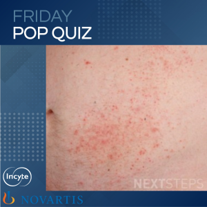
The correct answer is C. HLA-DQ2.
The image, along with the vignette, describes dermatitis herpetiformis (DH). The clinical presentation is very pruritic vesicles that are grouped and located symmetrically, especially on extensor surfaces. Overlying erosions can be seen due to the intense pruritus, and it can also present as papules, urticarial lesions, tense bullae, or polymorphous lesions. This rash can have cyclic exacerbations and is associated with Celiac disease. Histopathology, including direct immunofluorescence (DIF), helps aid in diagnosis – suprapapillary, neutrophil-rich, vesicles with a granular pattern on DIF with IgA>C3 along the dermal papillae. ELISA testing can also help in diagnosis, which will show antibodies against transglutaminase 3. HLA-DQ2 is commonly associated with DH, and treatment includes gluten-free diet, dapsone, and sulfapyridine.
HLA-Cw6 is most linked to psoriasis.
HLA-B51 is most linked to Behcet’s disease.
HLA-B27 is most linked to psoriatic arthritis or Reiter’s syndrome.
HLA-DR3 is most linked to Lupus (SCLE + SLE) and pemphigoid gestationis.
References:
Medical Dermatology chapter 2.3 Bullous and Vesicular Dermatosis, DIR Review book page 129
Brought to you by our brand partner
