
The correct answer is A. Transposition.
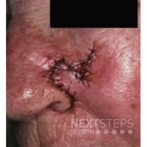
This is a rhombic flap which is a transposition flap. Transposition flaps are transposed over normal skin. Remember that a rhombic flaps end result can mimic a “question mark-like” shape. Here the tissue was transposed from the nasofacial sulcus to be used over the nasal sidewall and alar sidewall. The secondary defect created at the nasofacial sulcus would have been closed first – the “key-stitch” closes the secondary defect, which off-loads the flap to rest in the primary defect. The advantage of transposition flaps is the recruitment of tissue laxity from nearby basins and changing the primary vector of tension. Examples of transposition flaps: Rhombic, Bilobed, Nasolabial transposition, etc.
Advancement flaps (ex: O-T, A-T, O-U, O-H flaps) recruit nearby tissue and advance the tissue into the primary defect. In essence these are burrows triangle displacement flaps as the burrows triangles are taken in more cosmetically favorable locations or in areas of increased tissue laxity. This flap may appear to be an O-U flap but it is not.
Rotation flaps are similar to advancement flaps which displace the burrows triangle to a more favorable location or in an area of tissue laxity to recruit tissue to the primary defect. The main difference in the main plane of movement being rotational rather than in a straight line. Rotation and advancement flaps can both have vectors of rotation and advancement – they are named by their primary vector of movement. This is not a rotational flap.
This is not a linear closure. A linear closure is a standard closure where a burrows triangle is taken from either side of the defect and closed in a line in order to avoid standing cones or dog-ears.
This is not an interpolation flap. An interpolation flap is utilized to recruit tissue from a distant reservoir. They are placed into the primary defect and allowed to integrate with the primary defect site to allow maturation of blood supply there. A pedicle is left in place from the donor site which is then separated at around 3 weeks time.
References:
In Bolognia, J., In Schaffer, J. V., & In Cerroni, L. (2018). Dermatology.