
The correct answer is E. Langerin.
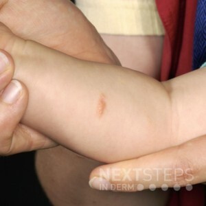
This is Hashimoto-Pritzker disease, benign single system Langerhans cell histiocytosis. It is characterized by congenital self-healing papulonodules that are red-brown color. It is composed of Langerhans cells which are langerin positive and CD1a positive and CD68 negative.
The appearance of the rash or X-rays and scans of internal organs may give the doctor a clue to the diagnosis. A skin biopsy of the affected area is usually needed to confirm the diagnosis. The biopsy findings do not give any indication of the likely course of the disease.
The diagnosis may be obvious when the biopsy tissue is looked at under a microscope, but often special stains are needed to clearly identify the type of histiocytosis.
When histiocytosis is diagnosed in one organ (eg, the skin or bone) other organs such as the blood, lungs, liver, and kidneys are checked to determine whether they are also affected by histiocytosis.
References:
Rook’s Textbook of Dermatology (7th Edition 2004), Chap 52, A Chu
Bolognia / Jorizzo / Rapini Dermatology Mosby publishing 2003, Chap 91 by Terry Barrett and CME Goodman.
Diamond EL, Subbiah V, Lockhart AC et al. Vemurafenib for BRAF V600-Mutant Erdheim-Chester Disease and Langerhans Cell Histiocytosis: Analysis of Data From the Histology-Independent, Phase 2, Open-label VE-BASKET Study. JAMA Oncol. 2018 Mar 1;4(3):384-388. doi: 10.1001/jamaoncol.2017.5029. PubMed PMID: 29188284; PubMed Central PMCID: PMC5844839. PubMed.
Brought to you by our Brand Partner
![]()