
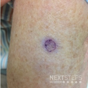
Answer: E. Parakeratosis in a column over focal dyskeratotic or vacuolated keratinocytes
This is an image of porokeratosis. While the center of the lesion can have variable histopathology (mild spongiosis or psoriasiform dermatitis), the raised border is represented histologically with a column of parakeratosis over often dyskeratotic or vacuolated keratinocytes. Regular elongated rete ridges are seen in psoriasis. Epidermal spongiosis with atypical lymphocytes spreading into the epidermis can be seen in mycosis fungoides. Perivascular lymphocytic infiltrate with prominent eosinophils can be seen with DRESS syndrome or other drug eruptions. Hyperplasia of the superficial epidermis can be seen in other benign epidermal tumors.
Don’t Agree? Tell us why in the comments section below.
Test your knowledge with custom quizzes at Derm In-Review or check out more pop quizzes.
Brought to you by our Brand Partner
![]()