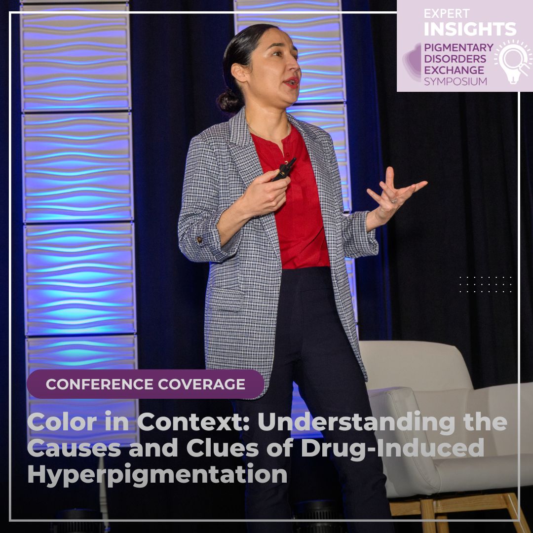
At the 2025 Pigmentary Disorders Exchange Symposium, Dr. Rebecca Vasquez, Associate Professor of Dermatology at UT Southwestern Medical Center, delivered an insightful presentation on drug-induced causes of hyperpigmentation. Her talk focused on the pathophysiology, clinical evaluation, and diagnostic tools used to differentiate these disorders from more common pigmentary conditions such as melasma or post-inflammatory hyperpigmentation.
Understanding the Epidemiology and Mechanisms
Drug-induced hyperpigmentation accounts for up to 20% of acquired pigmentation cases. While it can occur in all ages, races, and genders, individuals with deeply pigmented skin may experience more severe and longer-lasting discoloration. The mechanism of pigmentation varies by drug and can include melanin accumulation triggered by inflammation, direct deposition of the drug itself, stimulation of new pigment production (such as lipofuscin), or iron deposition.
Melanin accumulation is commonly seen with medications like antimalarials and may be exacerbated by sun exposure. Drugs such as silver can deposit directly into the skin, resulting in conditions like argyria. Some compounds trigger the formation of new pigments, while others, such as minocycline, result in iron deposition that contributes to a characteristic blue-gray or muddy discoloration.
Building the Diagnostic Framework
The foundation of evaluating suspected drug-induced pigmentation begins with a thorough history. Dr. Vasquez emphasized asking detailed questions about the onset of pigmentation, any recently introduced prescription or over-the-counter medications, herbal supplements, and whether symptoms changed with dosage adjustments or drug discontinuation. For example, pigmentation from minocycline is often dose-dependent and may worsen with higher cumulative exposure. Certain medications, such as antimalarials, can cause pigmentation that is aggravated by ultraviolet light or trauma.
To help determine causality, Dr. Vasquez recommended using the adverse drug reaction probability scale. This 10-question tool assigns a score to determine the likelihood that a drug caused the reaction, with a score above nine considered definitive.
On physical examination, clues such as the distribution of pigmentation, involvement of mucosa or nails, and specific coloration patterns can offer insights into the underlying mechanism. Brown tones typically indicate epidermal involvement, while blue-gray hues suggest dermal deposition. Mixed pigmentation, often seen in drug-induced cases, may appear as a combination of dark brown and steel-gray. The affected anatomical layers can further narrow the differential: epidermal pigmentation is often associated with lentigines and café-au-lait macules, while dermal pigmentation may suggest conditions such as lichen planus pigmentosus or erythema dyschromicum perstans.
Common Culprits and Clinical Clues
Hydroquinone: The Risk of Exogenous Ochronosis
Though widely used for skin lightening, prolonged or inappropriate use of hydroquinone can paradoxically result in exogenous ochronosis. This condition has been reported across all skin types but appears most frequently in Black patients, followed by Hispanic and Asian populations. A systematic review of 126 patients found an average duration of hydroquinone use of five years prior to diagnosis. The cheeks are most commonly affected (68%), followed by the forehead (24%) and temples (20%).
Clinicians should suspect ochronosis when patients report worsening of melasma despite treatment or describe improvement followed by darkening. Early signs include a mottled mix of erythema and blue-black hyperpigmentation, while late-stage findings consist of dark brown to black discoloration with “caviar-like” papules. The gold standard for diagnosis is histopathology, which reveals banana-shaped ochre fibers in the papillary dermis. However, dermoscopy can be a valuable noninvasive tool, revealing curvilinear, worm-like brown to black structures that may obviate the need for biopsy.
Antimalarials: Delayed but Distinctive Pigment Changes
Drugs such as chloroquine, hydroxychloroquine, and quinacrine—commonly used for autoimmune conditions like lupus—can lead to delayed-onset hyperpigmentation in 25–35% of patients, typically after a cumulative dose of 450 grams. Clinical presentation involves blue-gray pigmentation affecting the anterior shins, face, forearms, nail beds, and oral mucosa. These changes may be exacerbated by trauma or anticoagulant use, both of which contribute to hemosiderin deposition that stimulates melanogenesis. Histologic evaluation reveals both iron and melanin deposits in the dermis. Although some improvement may occur with discontinuation, resolution is often incomplete.
Minocycline: A Spectrum of Pigment Patterns
Minocycline, frequently prescribed for acne and rosacea, can cause progressive hyperpigmentation with long-term use. Pigment may deposit in the skin, sclera, teeth, and bones. Dr. Vasquez reviewed three distinct types of minocycline pigmentation:
-
- Type I: Blue-gray pigmentation in areas of inflammation or scarring, such as acne lesions
- Type II: Similar discoloration on the shins and forearms
- Type III: A diffuse, muddy brown pigmentation in sun-exposed areas
Types I and II may fade after discontinuation, whereas Type III is often permanent. Histology typically reveals iron and melanin in the dermis.
Silver (Argyria): A Classic but Rare Presentation
Silver, still used topically for burns, cauterization, or marketed in supplements as “immune boosters”, can lead to a condition known as argyria. This appears as slate-gray or metallic blue pigmentation on sun-exposed areas and may also affect the nails, oral mucosa, and sclera. Argyria can occur after years of ingestion or topical application. Histologic examination reveals black-brown granules within vessel walls and eccrine glands. Under polarized light, these deposits exhibit a “starry-sky” appearance.
Kratom: A Rising Herbal Offender
Kratom is a herbal supplement with opioid and stimulant-like properties that has gained popularity for pain relief in recent years. It can lead to generalized blue-gray hyperpigmentation, most notably in sun-exposed areas. Interestingly, the pigmentation tends to stop at the proximal interphalangeal joints, creating a sharp visual cutoff. Histopathology shows pigment deposition within macrophages and around blood vessels, which stains positively with Fontana-Masson. The pigmentation may improve with discontinuation, but clinical outcomes are variable.
In conclusion, hyperpigmentation is a common dermatologic concern, but not all pigmentation is melasma or post-inflammatory. Drug-induced causes require a careful history, attention to color and distribution, and the use of diagnostic aids like dermoscopy and histology. Dr. Vasquez’s lecture underscores the importance of recognizing patterns associated with specific agents from antimalarials and antibiotics to over-the-counter supplements like kratom and applying a systematic framework to improve diagnostic accuracy and patient care.
Did you enjoy this article? You can find more on hyperpigmentation here.

