
Friday Pop Quiz 7/24/2026
 An infant is referred by their pediatrician for further evaluation of seborrheic dermatitis refractory to standard therapy. What is the most appropriate intervention?
A. Prescribe clobetasol solution
B. Obtain bacterial culture
C. Obtain fungal culture
D. Obtain complete blood count
E. Obtain skin biopsy
To find out the correct answer and read the explanation, click here. …
An infant is referred by their pediatrician for further evaluation of seborrheic dermatitis refractory to standard therapy. What is the most appropriate intervention?
A. Prescribe clobetasol solution
B. Obtain bacterial culture
C. Obtain fungal culture
D. Obtain complete blood count
E. Obtain skin biopsy
To find out the correct answer and read the explanation, click here. …
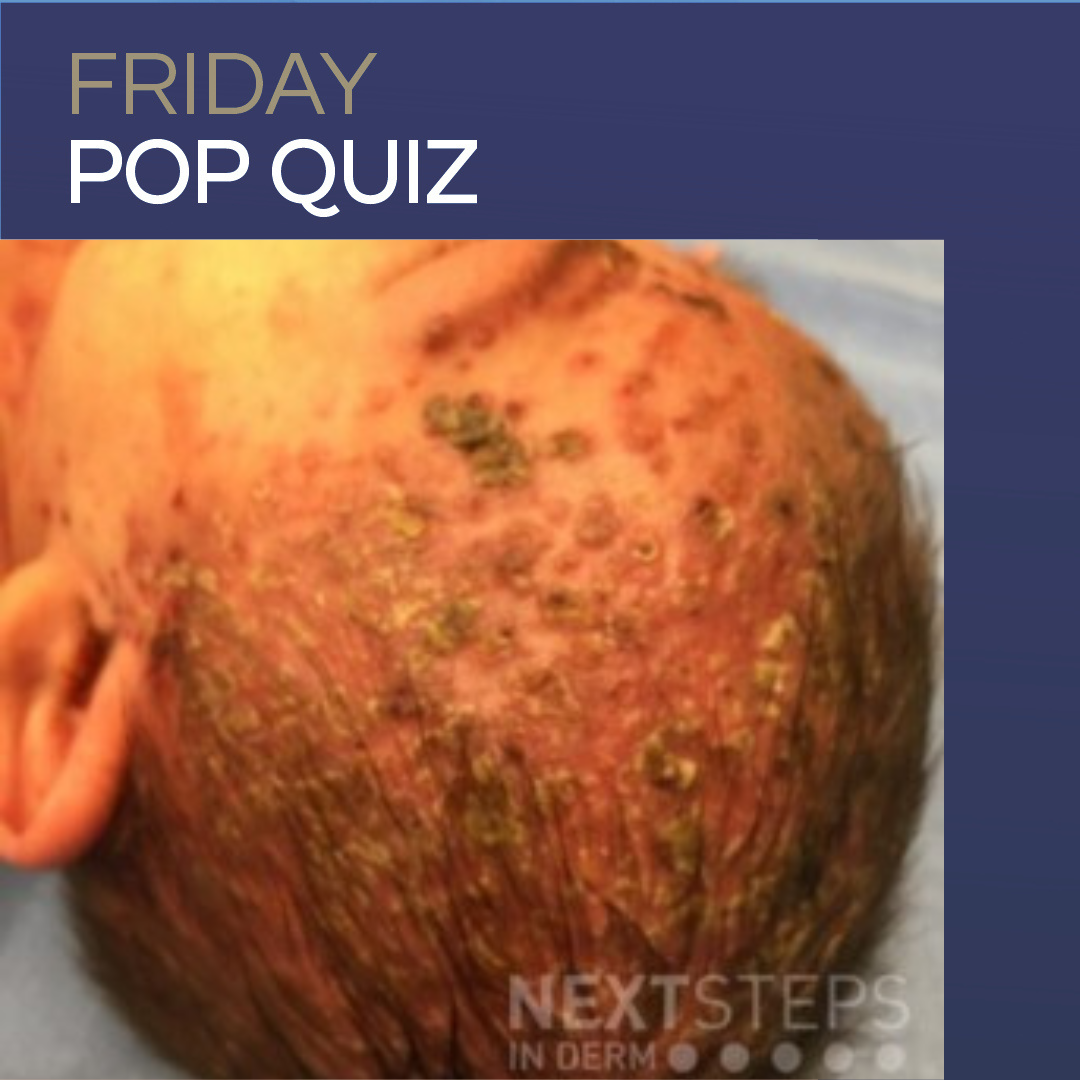
An infant is referred by their pediatrician for further evaluation of seborrheic dermatitis refractory to standard therapy. What is the most appropriate intervention?
A. Prescribe clobetasol solution
B. Obtain bacterial culture
C. Obtain fungal culture
D. Obtain complete blood count
E. Obtain skin biopsy
To find out the correct answer and read the explanation, click here. … 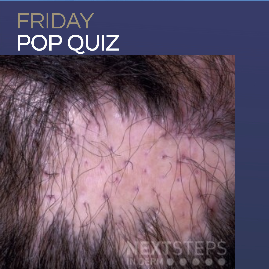 Which of the following is the most likely diagnosis in this 56-year-old woman with pruritic hair loss for 3 months?
A. Lichen planopilaris
B. Androgenetic alopecia
C. Central centrifugal cicatricial alopecia
D. Discoid lupus erythematosus
E. Folliculitis decalvans
F. Alopecia areata
To find out the correct answer and read the explanation, click here. …
Which of the following is the most likely diagnosis in this 56-year-old woman with pruritic hair loss for 3 months?
A. Lichen planopilaris
B. Androgenetic alopecia
C. Central centrifugal cicatricial alopecia
D. Discoid lupus erythematosus
E. Folliculitis decalvans
F. Alopecia areata
To find out the correct answer and read the explanation, click here. … 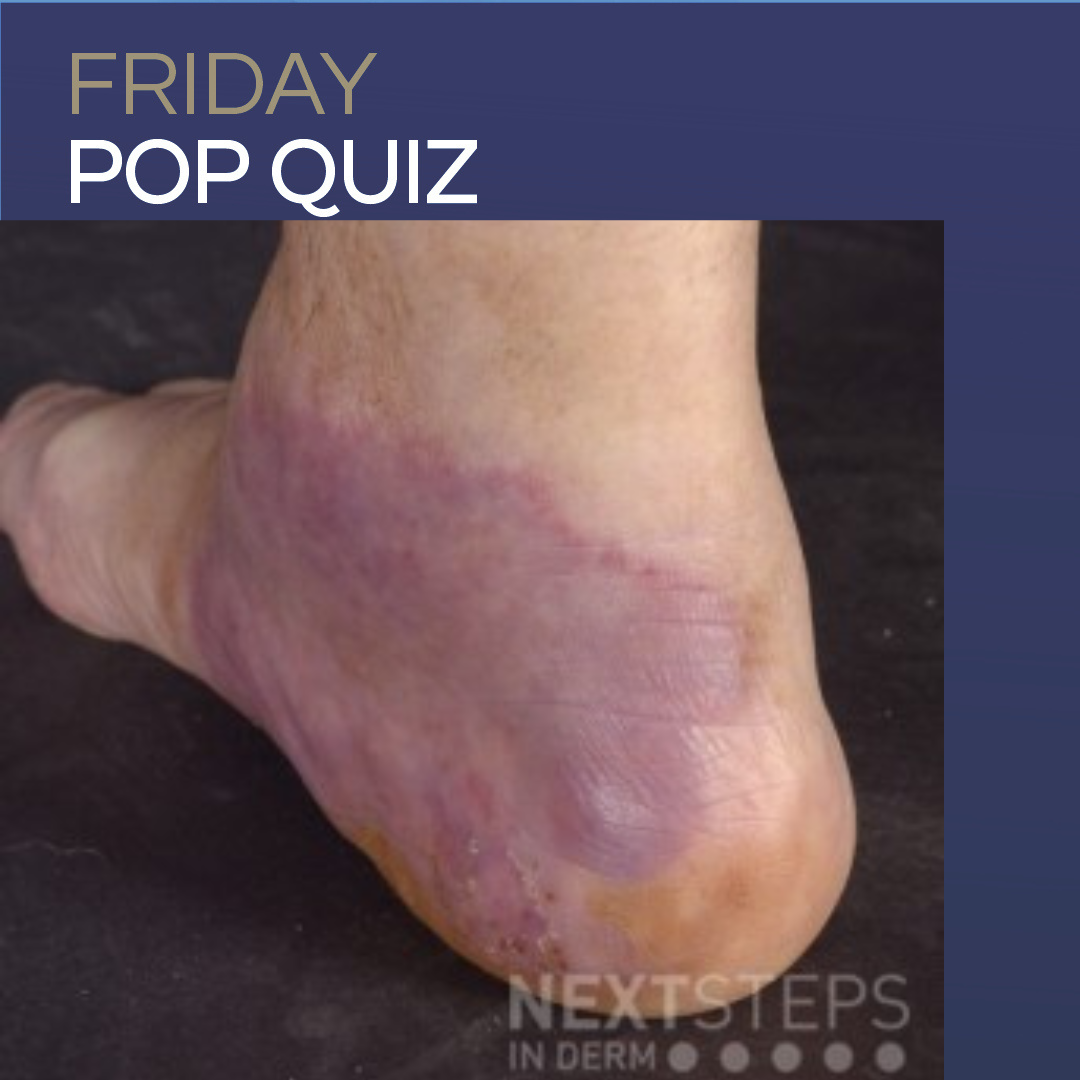 A 55 year old female presents with the following red-brown nodules on her elbows and similar appearing nodules and plaques on her ankles. She denies recent travel or any new sexual partners. She does note mild arthralgias associated with the rash. She is started on dapsone with some improvement. Which of the following is the best diagnosis?
A. Erythema induratum
B. Erythema elevatum diu …
A 55 year old female presents with the following red-brown nodules on her elbows and similar appearing nodules and plaques on her ankles. She denies recent travel or any new sexual partners. She does note mild arthralgias associated with the rash. She is started on dapsone with some improvement. Which of the following is the best diagnosis?
A. Erythema induratum
B. Erythema elevatum diu … 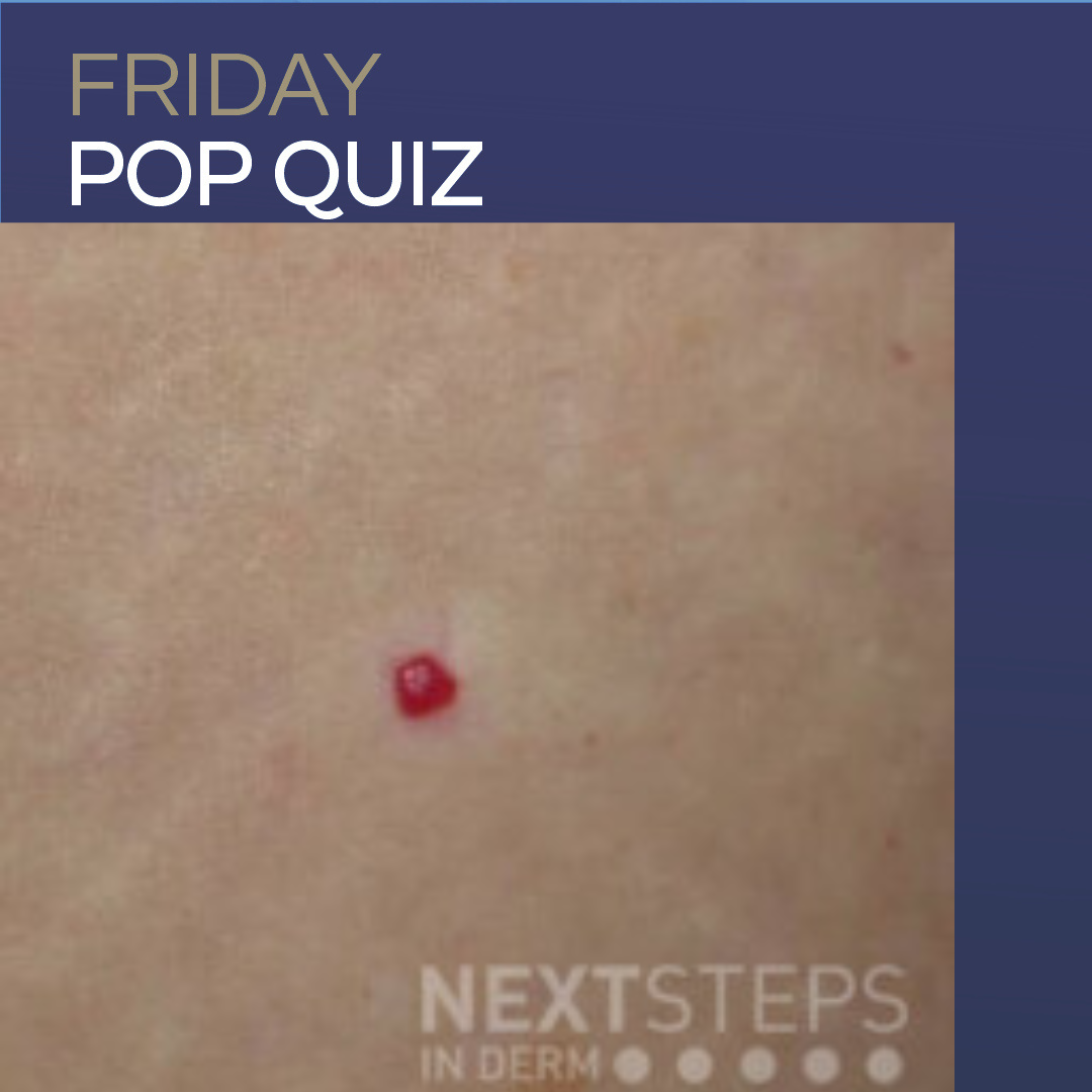 Which of the following wavelength and treatment settings are most appropriate for removal of the pictured lesion?
A. 1064 nm; 4 mm spot size, 110 J/cm2 fluence, and 30 ms pulse duration
B. 585 nm; 10 mm spot size, 10 J/cm2 fluence, and 20 ms pulse duration
C. 694 nm; 4 mm spot size, 3 J/cm2 fluence, and 20 ns pulse duration
D. 1064 nm; 10 mm spot size, 10 J/cm2, and 60 ms pulse dur …
Which of the following wavelength and treatment settings are most appropriate for removal of the pictured lesion?
A. 1064 nm; 4 mm spot size, 110 J/cm2 fluence, and 30 ms pulse duration
B. 585 nm; 10 mm spot size, 10 J/cm2 fluence, and 20 ms pulse duration
C. 694 nm; 4 mm spot size, 3 J/cm2 fluence, and 20 ns pulse duration
D. 1064 nm; 10 mm spot size, 10 J/cm2, and 60 ms pulse dur … 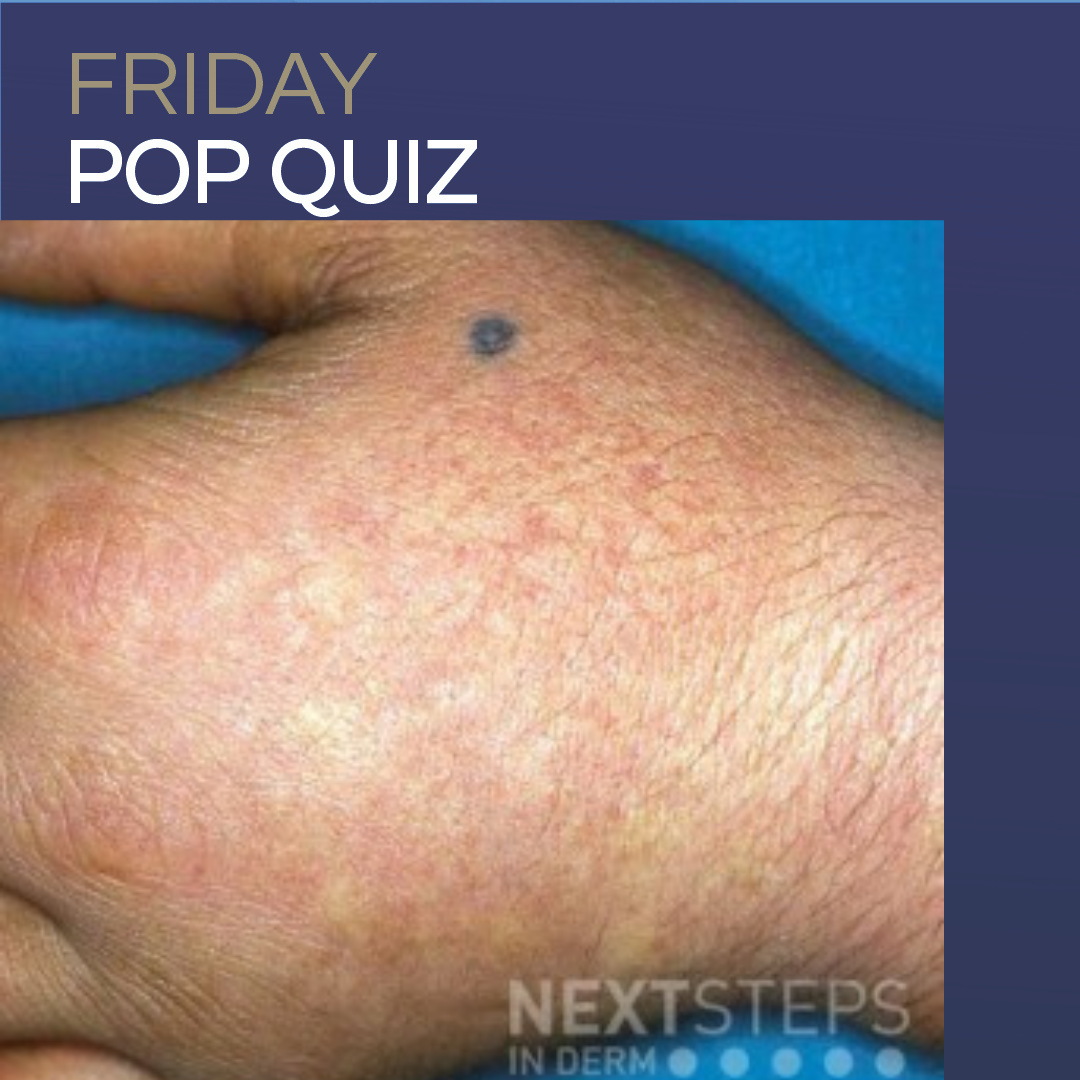 A 6-year-old male has painful edema and erythema of his posterior forearms, dorsal hands, and face several hours following sun exposure. He says a tingling and intense, painful burning sensation precede the rash when it occurs, starting immediately after sun exposure. These episodes recover in a few days. Episodes appear seasonal, with increased symptoms in the spring and summer months. Blood leve …