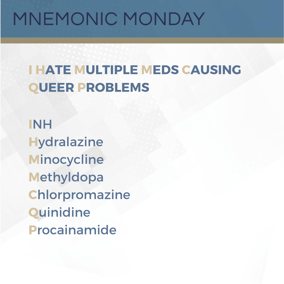
It’s Mnemonic Monday! I Hate Multiple Meds Causing Queer Problems
 On this Mnemonic Monday, we continue we continue with the topic of Systemic Lupus Erythematosus (SLE):
I HATE MULTIPLE MEDS CAUSING QUEER PROBLEMS
INH
Hydralazine
Minocycline
Methyldopa
Chlorpromazine
Quinidine
Procainamide
This mnemonic refers to drugs known to cause drug-induced Systemic Lupus Erythematosus (SLE).
Click HERE to print your mnemonic card.
Study More!
Need a r …
 Continue reading "It’s Mnemonic Monday! I Hate Multiple Meds Causing Queer Problems"


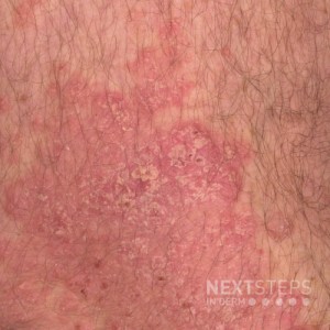
 The hyperproliferative epithelium of this mature psoriasis plaque is associated with increased expression of which keratin(s)?
A. K1, K10
B. K5, K14
C. K6, K16
D. K17
E. K2e
To find out the correct answer and read the explanation, click here.
Brought to you by our brand partner Derm In-Review. A product of SanovaWorks.
…
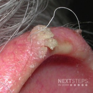
  Mutations in which gene would likely be found in the neoplastic cells of this lesion?
A. PATCH
B. p53
C. Fumarate hydratase
D. CREBBP
E. p63
To find out the correct answer and read the explanation, click here.
Brought to you by our brand partner Derm In-Review. A product of SanovaWorks.
…
  On this Mnemonic Monday, we challenge you to remember the criteria needed to diagnose Systemic Lupus Erythematosus (SLE) with the following mnemonic:
MD SOAP N HAIR
Malar erythema
Discoid LE
Serositis (carditis or pleurisy)
Oral ulcers
Arthritis, nonerosive
Photosensitivity
Neurologic (seizure or psychosis)
Hematologic disorder
ANA
Immunologic disorder
Renal
This mnemonic refers to …
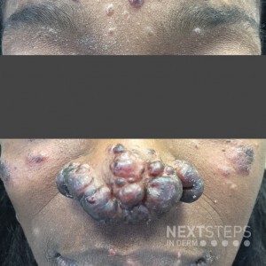
  Which extracutaneous organ is typically associated with this subtype of sarcoidosis?
A. Heart
B. Lungs
C. Kidneys
D. Eyes
E. Liver
To find out the correct answer and read the explanation, click here.
Brought to you by our brand partner Derm In-Review. A product of SanovaWorks.
…