
Key Tips for Dermatologists in Prescribing and Managing JAK Inhibitors
 Next Steps in Derm, in partnership with ODAC Dermatology, Aesthetic and Surgical Conference, interviewed Dr. Neal Bhatia (a board-certified Dermatologist who serves as Director of Clinical Dermatology at Therapeutics Clinical Research ) about the latest with JAK inhibitors. Watch as he summarizes the most recent trials, approvals, and developments. Plus why doctors shouldn't be afraid to dive in w …
Next Steps in Derm, in partnership with ODAC Dermatology, Aesthetic and Surgical Conference, interviewed Dr. Neal Bhatia (a board-certified Dermatologist who serves as Director of Clinical Dermatology at Therapeutics Clinical Research ) about the latest with JAK inhibitors. Watch as he summarizes the most recent trials, approvals, and developments. Plus why doctors shouldn't be afraid to dive in w … Continue reading "Key Tips for Dermatologists in Prescribing and Managing JAK Inhibitors"
 Data-Driven Dermatology Improves Patient Outcomes
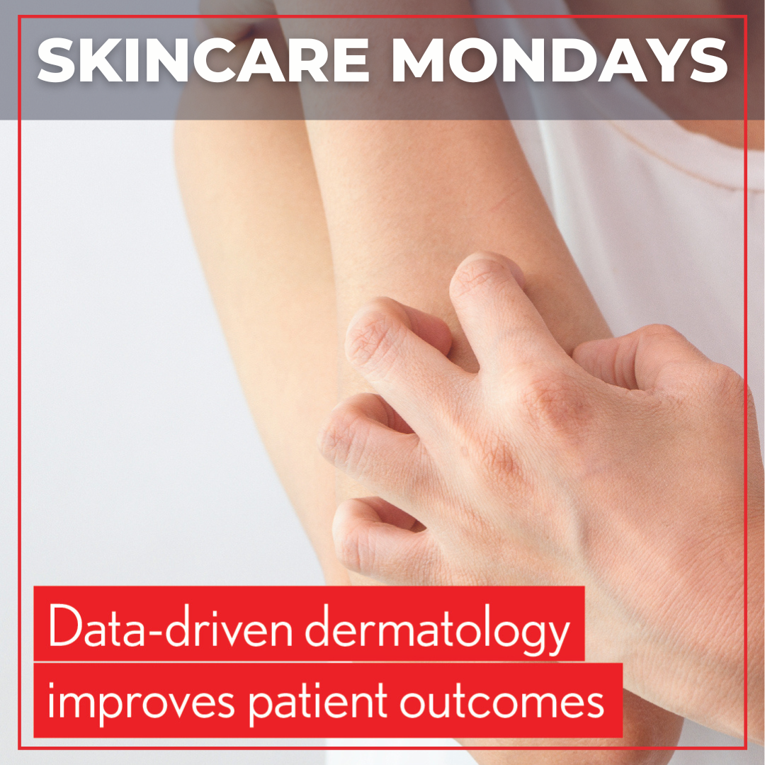
The use of real-world data and real-world evidence to inform health care decisions is increasing. While randomized controlled trials (RCTs) are still the gold standard for evidence-based medicine, the strict inclusion/exclusion criteria and tightly controlled conditions limit their generalizability to real-world clinical practice. Real worl …
Data-Driven Dermatology Improves Patient Outcomes
The use of real-world data and real-world evidence to inform health care decisions is increasing. While randomized controlled trials (RCTs) are still the gold standard for evidence-based medicine, the strict inclusion/exclusion criteria and tightly controlled conditions limit their generalizability to real-world clinical practice. Real worl … 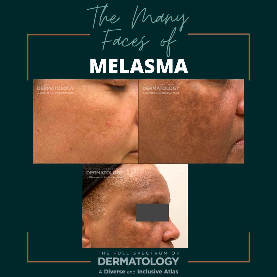 Our new series, “The Many Faces of”, showcases side-by-side images of some of the most commonly seen dermatology conditions in an array of skin tones and briefly highlight nuances in clinical presentation. All images featured in the series are part of The Full Spectrum of Dermatology: A Diverse and Inclusive Atlas, a resource developed by co-editors Misty Eleryan, MD, MS, and Adam Friedman, …
  Suncare is important for every skin tone. The risk of sunburn correlates with skin tone - not ethnicity.
Sunburn experiences differ across ethnicities.
An online survey of 3,597 adults who identified as White, Black, Hispanic and Asian showed sunburns occur across all ethnicities - even the darkest skin tones, but the experience is very different.1 Those who identified as White reported “ski …
Suncare is important for every skin tone. The risk of sunburn correlates with skin tone - not ethnicity.
Sunburn experiences differ across ethnicities.
An online survey of 3,597 adults who identified as White, Black, Hispanic and Asian showed sunburns occur across all ethnicities - even the darkest skin tones, but the experience is very different.1 Those who identified as White reported “ski … 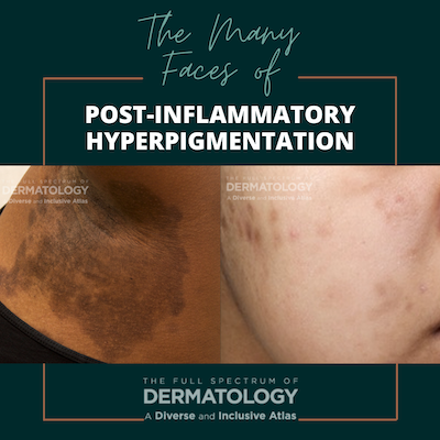 Our new series, “The Many Faces of”, showcases side-by-side images of some of the most commonly seen dermatology conditions in an array of skin tones and briefly highlight nuances in clinical presentation. All images featured in the series are part of The Full Spectrum of Dermatology: A Diverse and Inclusive Atlas, a resource developed by co-editors Misty Eleryan, MD, MS, and Adam Friedman, …