
Treatment of Hidradenitis Suppurativa in 2021 & Beyond
 At the ODAC 2021 Sneak Peek Symposium on Inflammatory Skin Diseases, expert faculty presented on the topics of acne, rosacea, atopic dermatitis, hidradenitis suppurativa, and psoriasis. If you missed the live symposium, Next Steps will be sharing highlights and a summary of each lecture over the course of the next few weeks. Today, Dr. Blari Allais shares an excellent recap of Dr. Joslyn Kirby's s …
At the ODAC 2021 Sneak Peek Symposium on Inflammatory Skin Diseases, expert faculty presented on the topics of acne, rosacea, atopic dermatitis, hidradenitis suppurativa, and psoriasis. If you missed the live symposium, Next Steps will be sharing highlights and a summary of each lecture over the course of the next few weeks. Today, Dr. Blari Allais shares an excellent recap of Dr. Joslyn Kirby's s … Continue reading "Treatment of Hidradenitis Suppurativa in 2021 & Beyond"
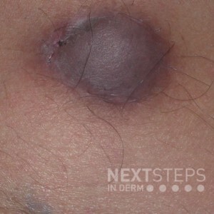 Histologically, this lesion shows plump, polygonal cells arranged in nests and fascicles with granular cytoplasm. Which immunohistochemical stain would be positive?
A. CD31
B. Synaptophysin
C. Factor XIIIa
D. S-100
E. CD34
To find out the correct answer and read the explanation, click here.
Brought to you by our brand partner Derm In-Review. A product of SanovaWorks.
…
Histologically, this lesion shows plump, polygonal cells arranged in nests and fascicles with granular cytoplasm. Which immunohistochemical stain would be positive?
A. CD31
B. Synaptophysin
C. Factor XIIIa
D. S-100
E. CD34
To find out the correct answer and read the explanation, click here.
Brought to you by our brand partner Derm In-Review. A product of SanovaWorks.
…  Management of alopecia in skin of color is challenging due to a paucity of research into its pathophysiology coupled with a poor understanding of the basic hair care practices in this patient population. For the patient, it is often associated with severe emotional distress. Unfortunately, many forms of hair loss are refractory to standard therapies.
At the 2020 Skin of Color Virtual Update, Dr …
Management of alopecia in skin of color is challenging due to a paucity of research into its pathophysiology coupled with a poor understanding of the basic hair care practices in this patient population. For the patient, it is often associated with severe emotional distress. Unfortunately, many forms of hair loss are refractory to standard therapies.
At the 2020 Skin of Color Virtual Update, Dr … 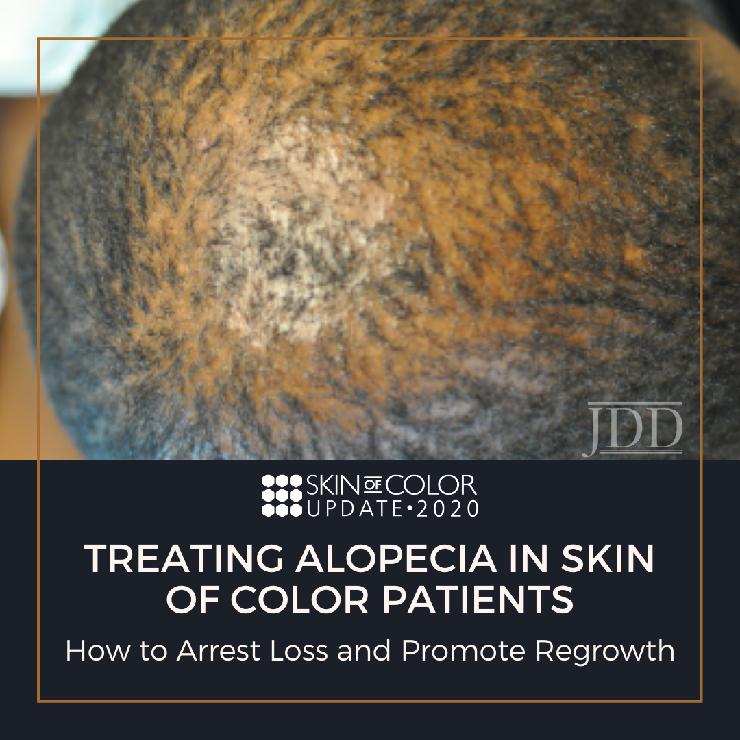 Can you improve your culturally-competent hair loss consultation? Would you like to grow your toolbox for comprehensive alopecia treatment? If so, you are in the right place!
Hair loss is a frequent concern for patients visiting the dermatologist, especially in patients with pigmented skin types. Kinky or coiled hair has an innate fragility that makes African Americans especially prone to hair …
Can you improve your culturally-competent hair loss consultation? Would you like to grow your toolbox for comprehensive alopecia treatment? If so, you are in the right place!
Hair loss is a frequent concern for patients visiting the dermatologist, especially in patients with pigmented skin types. Kinky or coiled hair has an innate fragility that makes African Americans especially prone to hair …  Women’s Health recently wrote an article about four skin conditions that are commonly misdiagnosed. Why do primary care providers sometimes misdiagnose common skin conditions? What conditions are challenging for dermatologists to diagnose? What steps can a dermatologist take to make a differential diagnosis?
For expert advice, I contacted Steve Daveluy, MD, FAAD, associate professor and pr …
Women’s Health recently wrote an article about four skin conditions that are commonly misdiagnosed. Why do primary care providers sometimes misdiagnose common skin conditions? What conditions are challenging for dermatologists to diagnose? What steps can a dermatologist take to make a differential diagnosis?
For expert advice, I contacted Steve Daveluy, MD, FAAD, associate professor and pr …