
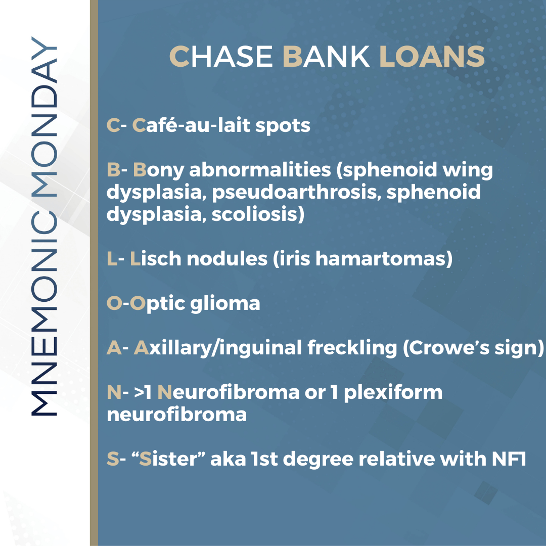
On this Mnemonic Monday, we challenge you to remember the major diagnostic criteria for neurofibromatosis type I with the following mnemonic:
Chase Bank LOANS
C– Café-au-lait spots
B– Bony abnormalities (sphenoid wing dysplasia, pseudoarthrosis, sphenoid dysplasia, scoliosis)
L– Lisch nodules (iris hamartomas)
O–Optic glioma
A– Axillary/inguinal freckling (Crowe’s sign)
N– >1 Neurofibroma or 1 plexiform neurofibroma
S– “Sister” aka 1st degree relative with NF1
This mnemonic refers to the major diagnostic criteria for neurofibromatosis type I. Two major diagnostic criteria are needed. The major diagnostic criteria include: 6 or more café au lait macules, axillary or inguinal freckling (Crowe’s sign), 2 or more neurofibromas or 1 plexiform neurofibroma, 2 or more Lisch nodules (iris hamartomas), optic glioma, bony lesions (pseudoarthrosis osseous lesions, sphenoid dysplasia, scoliosis), 1st-degree relative with NF-1.
Click HERE to download or print your mnemonic card.
Study More!
Need a refresher on neurofibromatosis? Check out pages 225-226 of your 2020 Derm In-Review Study Guide.
Don’t have a copy? Sign up for Derm In-Review and download the digital version here.
Further Reading
Check out the following case report published in the Journal of Drugs in Dermatology (JDD):
Eruptive Plexiform Schwannomas in a Child With Neurofibromatosis I
Introduction
Case Report
A nine-year-old male with a past medical history significant for a cataract at age two, with no known family history of neurocutaneous disorders, presented with a stumbling gait and hip pain in May 2010. Orthopedic examination revealed no traumatic cause and a neurological evaluation was undertaken. Two months later, in July of 2010, an MRI of his spine revealed a thoracic spine tumor. Within weeks, paralysis ensued. Surgical resection of the tumor revealed a meningioma at the level of T5/T6. On further examination, bilateral acoustic neuromas were discovered despite the patient having no hearing deficits. A diagnosis of Neurofibromatosis II was rendered at this time.


Test your knowledge!
Which of the following signs is not a criterion for the diagnosis of Neurofibromatosis type I?
A: Bilateral vestibular schwannomas
B: Axillary freckling
C: Optic gliomas
D: Greater than 5 café-au-lait macules
E: Sphenoid dysplasia
To find out the correct answer and read the explanation, click here.
Did you enjoy this mnemonic? You can find more here.
Brought to you by our brand partner Derm In-Review. A product of SanovaWorks.
![]()