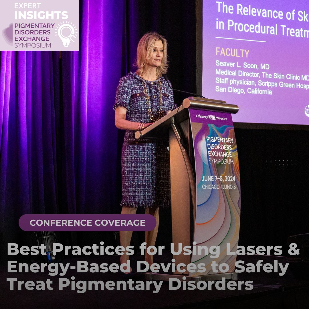
At the 2024 Pigmentary Disorder Exchange Symposium, Dr. Arielle Kauvar, an internationally renowned board-certified dermatologist, laser surgeon, and Mohs surgeon, delivered a lecture focused on best practices for safely treating pigmentary disorders using lasers and energy-based devices, particularly in skin of color. If you missed the lecture, do not fret! This summary reviews the key takeaways regarding device selection, risk mitigation, and the importance of patient management in procedural treatments.
Understanding the Risks in Skin of Color
Dr. Kauvar began by highlighting the critical need to treat all skin types safely and effectively, particularly in patients with skin of color, who are at a greater risk for pigmentary alterations and scarring. She pointed out that many providers are reluctant to use lasers and energy-based devices in this population due to the limited data and elevated risk posed by the higher melanin content in their skin. As the U.S. demographic shifts and more skin of color patients seek cosmetic treatments, it is increasingly important for providers to fully understand both the direct and indirect risks associated with using these devices in such populations.
-
- Direct risks arise from melanin/melanocyte damage or disruption of the dermal-epidermal junction.
- Occur with lasers in the visible light range (400-700nm) and with intense pulsed light (IPL) devices. These wavelengths are strongly absorbed by melanin, increasing the risk of hyperpigmentation and scarring.
- Indirect risks come from the effects of tissue heating, which stimulates melanogenesis, leading to post-inflammatory hyperpigmentation (PIH), particularly in Asian and African American populations. These patient populations also are known to have a higher risk of hypertrophic and keloidal scarring compared to white patients.1,2
- Bulk tissue heating can trigger inflammation and stimulate melanogenesis, resulting in hyperpigmentation. This is most notably observed with ablative lasers, both fractional and full beam, as well as with high-density coverage using non-ablative lasers. Additionally, high-fluence treatments with pulse lasers or pulse stacking, and even low-fluence treatments can deliver excessive energy to the skin if multiple passes (10 – 20) are performed, significantly increasing the risk of hyperpigmentation.
- Direct risks arise from melanin/melanocyte damage or disruption of the dermal-epidermal junction.
Device Selection for Skin of Color
Pigment-Specific Lasers
Nanosecond and picosecond lasers are ideal adjunct therapies for disorders like melasma due to their precision in targeting melanosomes and melanocytes without affecting surrounding tissue.
-
- Nanosecond lasers target larger structures such as melanocytes or melanophages using thermal energy.
- Nanosecond = a billionth of second
- Higher risk of tissue heating and PIH
- Picosecond lasers target melanosomes with photomechanical effects, reducing tissue heating and improving recovery.
- Picosecond= a trillionth of a second
- Minimal heating and risk of PIH
- Nanosecond lasers target larger structures such as melanocytes or melanophages using thermal energy.
Color-Blind Devices
Non-ablative fractional lasers, radiofrequency (RF) devices, and ultrasound energy are considered safer alternatives because they are not absorbed by melanin, pose minimal risk of bulk heating, and spare the epidermis.3-5
-
- High-intensity focused ultrasound (HIFU) creates coagulation zones at different depths, stimulating collagen production without damaging the epidermis. It has been shown to effectively treat wrinkles, lift skin, and improve skin laxity with low risk of hyperpigmentation.6
- Synchronous Ultrasound Parallel Beam (SUPERB) Technology: This novel system employs seven high-frequency synchronous ultrasound beams to heat the skin to temperatures between 60 and 70 degrees Celsius for four to five seconds. These beams create coagulation zones that are aligned parallel to the skin surface, rather than perpendicular. This technique heats a planar section of tissue, achieving extensive coverage at depths of 1.5 to 2 millimeters below the surface while completely sparing the epidermis. Each treatment results in heating 25% to 30% of the targeted tissue in a single plane. This thermal effect stimulates the production of collagen, elastin, and mucopolysaccharides, as evidenced by immunohistochemistry staining.7
- Monopolar, bipolar, and tripolar RF devices have been shown to effectively treat wrinkles, lift skin, and improve acne scars.8,9
Best Practices for Safe and Effective Treatment
Dr. Kauvar stressed that the safe use of energy-based devices in skin of color requires dedicating time to thoroughly evaluate each patient. One should conduct a comprehensive medical history and physical examination to assess risk factors like a predisposition to hyperpigmentation. Patient education is also crucial to set realistic expectations and inform them of potential risks.
A thorough understanding of laser-tissue interactions is key to safely targeting specific chromophores without causing collateral damage.10 Dr. Kauvar highlighted the three principles of selective photothermolysis:
-
- Wavelength: Choose a wavelength that will be absorbed by your target chromophore (i.e., melanin, blood, or water). Wavelength determines the depth of laser penetration and melanin absorption.
- Longer wavelengths (e.g., 1064nm Nd:YAG) penetrate deeper and are safer for dermal melanophages in skin of color.
- Shorter wavelengths (e.g., 532nm) are absorbed more superficially and pose higher risks in melanated skin.
- Pulse Duration: Choose a duration shorter than the thermal relaxation time to limit damage to the target without affecting surrounding tissues.
- Larger targets with longer thermal relaxation time require a longer pulse duration.
- Melanosome (0.1 micron) targeted by picosecond lasers
- Melanocyte or melanophage (10-80 microns) targeted by nanosecond lasers
- Larger targets with longer thermal relaxation time require a longer pulse duration.
- Wavelength: Choose a wavelength that will be absorbed by your target chromophore (i.e., melanin, blood, or water). Wavelength determines the depth of laser penetration and melanin absorption.
-
- Fluence (Energy Density): Should be sufficient to cause irreversible damage to the target while minimizing risk to surrounding tissues.
Dr. Kauvar also recommended using topical agents (i.e., topical steroids) both before and after the procedure to minimize inflammation and prevent melanocyte activation, thereby reducing the risk of PIH in skin color. Additionally, using bulk cooling between laser passes can help protect the epidermis and reduce heat buildup.
In conclusion, lasers offer greater potential for safety and efficacy in treating skin of color compared to non-selective destructive modalities like electrodesiccation or liquid nitrogen. Dr. Kauvar emphasized that safe and effective treatment requires selecting the appropriate device, conducting a thorough patient evaluation, and understanding laser-tissue interactions. However, there is a clear need for more clinical trials and data on the use of lasers and energy-based devices in skin of color. Collaboration between device manufacturers and clinicians is essential to advance technology and safety protocols. With ongoing industry innovations and research partnerships, procedural treatments for diverse skin types can be further optimized to enhance both safety and efficacy.
References
-
- Soltani, A. M., Francis, C. S., Motamed, A., Karatsonyi, A. L., Hammoudeh, J. A., Sanchez-Lara, P. A., Reinisch, J. F., & Urata, M. M. (2012). Hypertrophic scarring in cleft lip repair: a comparison of incidence among ethnic groups. Clinical epidemiology, 4, 187–191. https://doi.org/10.2147/CLEP.S31119
- Chike-Obi, C. J., Cole, P. D., & Brissett, A. E. (2009). Keloids: pathogenesis, clinical features, and management. Seminars in plastic surgery, 23(3), 178–184. https://doi.org/10.1055/s-0029-1224797
- Kauvar A. N. (2012). The evolution of melasma therapy: targeting melanosomes using low-fluence Q-switched neodymium-doped yttrium aluminium garnet lasers. Seminars in cutaneous medicine and surgery, 31(2), 126–132. https://doi.org/10.1016/j.sder.2012.02.002
- Friedmann, D. P., Tzu, J. E., Kauvar, A. N., & Goldman, M. P. (2016). Treatment of facial photodamage and rhytides using a novel 1,565 nm non-ablative fractional erbium-doped fiber laser. Lasers in surgery and medicine, 48(2), 174–180. https://doi.org/10.1002/lsm.22461
- Kauvar, A. N. B., & Gershonowitz, A. (2022). Clinical and histologic evaluation of a fractional radiofrequency treatment of wrinkles and skin texture with novel 1-mm long ultra-thin electrode pins. Lasers in surgery and medicine, 54(1), 54–61. https://doi.org/10.1002/lsm.23452
- Fabi, S. G., & Goldman, M. P. (2014). Retrospective evaluation of micro-focused ultrasound for lifting and tightening the face and neck. Dermatologic surgery : official publication for American Society for Dermatologic Surgery [et al.], 40(5), 569–575. https://doi.org/10.1111/dsu.12471
- Wang, J. V., Ferzli, G., Jeon, H., Geronemus, R. G., & Kauvar, A. (2021). Efficacy and Safety of High-Intensity, High-Frequency, Parallel Ultrasound Beams for Fine Lines and Wrinkles. Dermatologic surgery : official publication for American Society for Dermatologic Surgery [et al.], 47(12), 1585–1589. https://doi.org/10.1097/DSS.0000000000003208
- Syder, N. C., Chen, A., & Elbuluk, N. (2023). Radiofrequency and Radiofrequency Microneedling in Skin of Color: A Review of Usage, Safety, and Efficacy. Dermatologic surgery : official publication for American Society for Dermatologic Surgery [et al.], 49(5), 489–493. https://doi.org/10.1097/DSS.0000000000003733
- Gold, M. H., & Biron, J. (2020). Improvement of wrinkles and skin tightening using TriPollar® radiofrequency with Dynamic Muscle Activation (DMA™). Journal of cosmetic dermatology, 19(9), 2282–2287. https://doi.org/10.1111/jocd.13620
- Kauvar, A. N. B., Kubicki, S. L., Suggs, A. K., & Friedman, P. M. (2020). Laser Therapy of Traumatic and Surgical Scars and an Algorithm for Their Treatment. Lasers in surgery and medicine, 52(2), 125–136. https://doi.org/10.1002/lsm.23171
This information was presented by Dr. Arielle Kauvar during the 2024 Pigmentary Disorders Exchange Symposium. The above session highlights were written and compiled by Dr. Sarah Millan.
Did you enjoy this article? You can find more here.

