> Skin of Color

Widespread Skin Necrosis Secondary to Gemcitabine Therapy

INTRODUCTION

Cutaneous side effects are relatively common in chemotherapy but vary in frequency and severity depending on the medication, dose, duration, pharmacokinetics, and pharmacodynamics. Gemcitabine, a pyrimidine nucleoside analogue, is a common oncologic agent used in the treatment of a variety of malignancies such as cancers of the head and neck, lung cancer, breast cancer, pancreatic cancer, non-Hodgkin lymphoma, and CTCL. Common adverse events include nausea, vomiting, and myelosuppression. Severe side effects such as hemolytic uremic syndrome and thrombotic thrombocytopenic purpura are uncommon. Herein we present an unusual case of widespread skin necrosis mimicking toxic epidermal necrolysis but lacking mucosal involvement in a patient receiving gemcitabine therapy for cutaneous T-cell lymphoma.


CASE REPORT

A 45-year-old woman with past medical history of Stage IIIA CTCL treated previously with topical nitrogen mustard, bexarotene, interferon alpha 2b, romidepsin, pembrolizumab, and brentuximab vedotin was admitted to a community hospital with erythroderma, skin erosions, and intermittent fever. Of note, the patient had not received systemic chemotherapy in the 8 weeks prior to this acute presentation. Upon admission, she was diagnosed with otitis externa and herpes simplex virus (HSV) of the face and was treated with vancomycin, meropenem, micafungin, and acyclovir, which resulted in defervescence. Six days later, the patient received her first dose of gemcitabine, which initially resulted in clearing of her skin. The patient received a second dose of intravenous gemcitabine 1000 mg/m2 one week later. Two days after the second dose of gemcitabine, she developed cytopenia-requiring filgrastim (Neupogen) and generalized full-thickness necrosis of the neck, trunk, and extremities without mucosal involvement (Figure 1A-C). Gemcitabine was discontinued and the patient was transferred to our facility for wound care and further treatment.


Upon admission to our hospital, she was noted to be afebrile. Workup included basic metabolic panel and liver function tests, which were within normal limits. She was noted to be anemic with hemoglobin of 6.5 g/dL and was given a transfusion of 1 unit of packed red blood cells (PRBC). The patient’s albumin was 1.4 and nutrition was optimized. Other relevant laboratory data included a CD4/CD8 ratio of 1.26, Sezary count of 0%, and HTLV I/II were negative. She was placed on combination medical therapy with intravenous methylprednisolone 40 mg every 8 hours, intravenous acyclovir 10 mg/kg three times a day, intravenous vancomycin 1 g every 8 hours, and intravenous meropenem 1 g every 8 hours. Additionally, oral acitretin 25 mg was administered every other day and increased to 25 mg daily on day 19. Wound care included nonstick sheets, dressings with 0.5% silver nitrate and sterile water every 8 hours changed every 3-4 days with bedside monitored anesthesia care (MAC). The patient remained afebrile and blood cultures were negative, therefore antibiotic therapy was discontinued on hospital day five and acyclovir was changed from intravenous to oral. Repeat wound cultures were negative for HSV but were positive for candida parapsilosis and mixed gram-positive flora indicating normal skin flora contaminants.


Histopathological examination of skin biopsies revealed residual cutaneous T-cell lymphoma with CD30-negative large cell transformation. The epidermis showed chemotherapy-related changes (Figure 2A-H). No viable epidermis was identified. Gram stain was negative for bacteria. HSV type 1 and type 2 immunostains were both negative.
Skin Necrosis
FIGURE 1. Clinical images. (A-C) Desquamation of the neck, trunk, and extremities on initial presentation. (D-F) Day 19 of hospital stay following wound care and treatment.

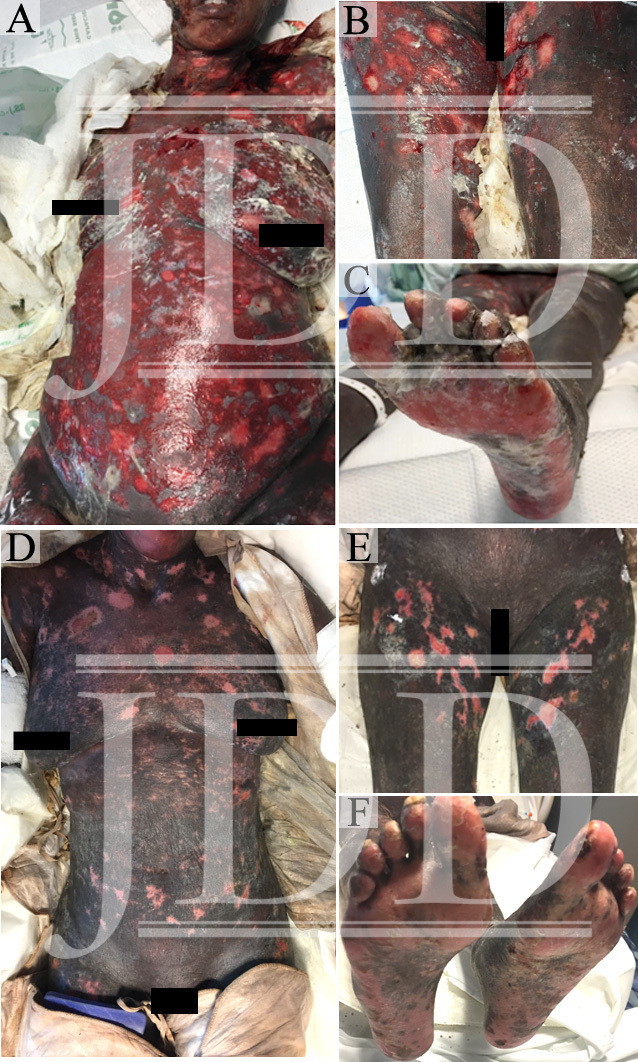
The patient’s skin continued to epithelialize and she was ultimately treated with extracorporeal photophoresis, two consecutive sessions every two weeks, acitretin 25 mg daily, and a systemic corticosteroid taper. She was discharged from hospital on day 32 and transferred to an inpatient rehab facility. The patient’s wound recovery can be seen at hospital day 19 (Figure 1D-F).

Skin Necrosis
FIGURE 2. Representative images of histopathologic features. (A-C) The dermis shows residual lymphoma cells. The epidermis shows chemotherapy effect (H&E, X400 and X200). (A-C) Immunohistochemistry results that the majority of the lichenoid infiltrates are CD3+ T-cells with an increased CD4 vs CD8 ratio. A minimal loss of CD7 expression is noted. Large cells are less than 25% of entire infiltrates, which shows CD30 expression in a few large cells. There are a large number of CD8+ T-cells and a minimal loss of CD7 expression.

DISCUSSION

Gemcitabine is a fluorine-substituted deoxycytidine analogue that is cell-cycle specific to S-phase and G1/S-phases. Used in a variety of oncologic settings, gemcitabine is metabolized by nucleoside kinases into two active metabolites: gemcitabine triphosphate (dFdCTP) and gemcitabine diphosphate (dFdCDP). Its antitumor effects are the result of inhibition of ribonucleotide reductase by gemcitabine diphosphate, inhibition of DNA polymerase by gemcitabine triphosphate, and incorporation of gemcitabine triphosphate into DNA resulting in chain termination.
Common toxicities include myelosuppression, nausea, vomiting, diarrhea, shortness of breath, and peripheral edema.1,2Common dermatologic reactions associated with gemcitabine include alopecia, mild skin rash, and mucositis. Rare reports of other cutaneous reactions are noted to occur including lower extremity skin necrosis,3lower extremity pseudocellulitis4, scleroderma-like changes2, cutaneous vasculitis,5,6 thrombotic microangiopathy,7-10 digital ischemia,11,12 linear immunoglobulin A bullous dermatitis (LABD),13 erysipeloid,14,15 and radiation recall dermatitis.16-18 Full-body skin necrosis is a rare phenomenon. One case of Stevens-Johnson syndrome (SJS) has been reported in a patient undergoing radiotherapy and gemcitabine19. In addition, there is one case report of toxic epidermal necrolysis (TEN) reported in a patient on gemcitabine for transition cell carcinoma of the bladder; however, this was a clinical diagnosis where biopsy was not performed.20 Stevens-Johnson syndrome and toxic epidermal necrolysis have been associated with alkylating agents, methotrexate, L-asparaginase, doxorubicin, docetaxel, and thalidomide.20 Cytarabine is another deoxycytidine analog chemotherapeutic agent closely related to gemcitabine in structure and mechanism of action. Cytarabine has been implicated in SJS/TEN in a number of cases; however, gemcitabine has not.20-23


Gemcitabine has been shown to be efficacious in CTCL and it is usually well tolerated24,25; however, a multicenter study of 23 cases noted two cases of bullous and erosive dermatitis among other side effects including hemolytic-uremic syndrome. CTCL, depending on staging, can be treated with topical nitrogen mustard, psoralen, and ultraviolet A radiation (PUVA), systemic retinoids, methotrexate, gemcitabine, pegylated liposomal doxorubicin, interferon-alpha 2a, romidepsin (histone deacetylase inhibitor), brentuximab vedotin (anti-CD30 monoclonal antibody conjugated with monomethyl auristatin E).25,26 Our patient had been treated with a number of agents over the past ten years without cutaneous reactions. However, when hospitalized and placed on gemcitabine for two cycles the patient developed severe cutaneous toxicity two days after the second dose. The patient improved with discontinuation of gemcitabine and meticulous wound care.


CONCLUSION

The pathogenesis of gemcitabine-induced cutaneous toxicities remains unclear. While cutaneous adverse effects have been reported with use of gemcitabine, these usually include skin eruptions, injection site reactions and alopecia. To the best of our knowledge, this is the first reported case of a TEN-like reaction in a patient receiving gemcitabine therapy for cutaneous T-cell lymphoma.


REFERENCES

    1. Aapro MS, Martin C, Hatty S. Gemcitabine–a safety review. Anticancer Drugs. 1998;9(3):191-201.
    2. Bessis D, Guillot B, Legouffe E, Guilhou JJ. Gemcitabine-associated scleroderma-like changes of the lower extremities. J Am Acad Dermatol. 2004;51(2 Suppl):S73-76.
    3. D’Epiro S, Salvi M, Mattozzi C, et al. Gemcitabine-induced extensive skin necrosis. Case Rep Med. 2012;2012:831616.
    4. Korniyenko A, Lozada J, Ranade A, Sandhu G. Recurrent lower extremity pseudocellulitis. Am J Ther. 2012;19(4):e141-142.
    5. Contreras-Steyls M, Lopez-Navarro N, Gallego E, Moyano B, Estrada D, Herrera E. Gemcitabine therapy-associated cutaneous vasculitis with a polyarteritis nodosa-like pattern. Int J Dermatol. 2013;52(8):1029-1031.
    6. Corella F, Dalmau J, Roe E, Garcia-Navarro X, Alomar A. Cutaneous vasculitis associated with gemcitabine therapy. Clin Exp Dermatol. 2009;34(1):97-99.
    7. Lai-Tiong F, Duval Y, Krabansky F. Gemcitabine-associated thrombotic microangiopathy in a patient with lung cancer: A case report. Oncol Lett. 2017;13(3):1201-1203.
    8. Turner JL, Reardon J, Bekaii-Saab T, Cataland SR, Arango MJ. Gemcitabine-Associated Thrombotic Microangiopathy: Response to Complement Inhibition and Reinitiation of Gemcitabine. Clin Colorectal Cancer. 2016; S1533-0028(16):30178-5.
    9. Saif MW, Xyla V, Makrilia N, Bliziotis I, Syrigos K. Thrombotic microangiopathy associated with gemcitabine: rare but real. Expert Opin Drug Saf. 2009;8(3):257-260.
    10. Humphreys BD, Sharman JP, Henderson JM, et al. Gemcitabine-associated thrombotic microangiopathy. Cancer. 2004;100(12):2664-2670.
    11. Kuhar CG, Mesti T, Zakotnik B. Digital ischemic events related to gemcitabine: Report of two cases and a systematic review. Radiol Oncol. 2010;44(4):257-261.
    12. Holstein A, Batge R, Egberts EH. Gemcitabine induced digital ischaemia and necrosis. Eur J Cancer Care (Engl). 2010;19(3):408-409.
    13. del Pozo J, Martinez W, Yebra-Pimentel MT, Almagro M, Pena-Penabad C, Fonseca E. Linear immunoglobulin A bullous dermatosis induced by gemcitabine. Ann Pharmacother. 2001;35(7-8):891-893.
    14. Ruiz-Casado A, Gutierrez D, Juez I. Erysipeloid rash: A rare adverse event induced by gemcitabine. J Cancer Res Ther. 2015;11(4):1024.
    15. Zustovich F, Pavei P, Cartei G. Erysipeloid skin toxicity induced by gemcitabine. J Eur Acad Dermatol Venereol. 2006;20(6):757-758.
    16. Schwartz BM, Khuntia D, Kennedy AW, Markman M. Gemcitabine-induced radiation recall dermatitis following whole pelvic radiation therapy. Gynecol Oncol. 2003;91(2):421-422.
    17. Burstein HJ. Side effects of chemotherapy. Case 1. Radiation recall dermatitis from gemcitabine. J Clin Oncol. 2000;18(3):693-694.
    18. Bar-Sela G, Beny A, Bergman R, Kuten A. Gemcitabine-induced radiation recall dermatitis: case report. Tumori. 2001;87(6):428-430.
    19. Sommers KR, Kong KM, Bui DT, Fruehauf JP, Holcombe RF. Stevens-Johnson syndrome/toxic epidermal necrolysis in a patient receiving concurrent radiation and gemcitabine. Anticancer Drugs. 2003;14(8):659-662.
    20. Mermershtain W, Cohen AD, Lazarev I, Grunwald M, Ariad S. Toxic epidermal necrolysis associated with gemcitabine therapy in a patient with metastatic transitional cell carcinoma of the bladder. J Chemother. 2003;15(5):510-511.
    21. Calista D, Landi C. Cytarabine-induced acral erythema: a localized form of toxic epidermal necrolysis? J Eur Acad Dermatol Venereol. 1998;10(3):274-275
    22. Ozkan A, Apak H, Celkan T, Yuksel L, Yildiz I. Toxic epidermal necrolysis after the use of high-dose cytosine arabinoside. Pediatr Dermatol. 2001;18(1):38-40.
    23. Figueiredo MS, Yamamoto M, Kerbauy J. [Toxic epidermal necrolysis after the use of intermediate dose of cytosine arabinoside]. Rev Assoc Med Bras (1992). 1998;44(1):53-55.
    24. Jidar K, Ingen-Housz-Oro S, Beylot-Barry M, et al. Gemcitabine treatment in cutaneous T-cell lymphoma: a multicentre study of 23 cases. Br J Dermatol. 2009;161(3):660-663.
    25. Network NCC. Mycosis Fungoides/Sezary Syndrome (Version 1.2016). https://www.nccn.org/professionals/physician_gls/pdf/t-cell.pdf. Accessed August 8, 2017.
    26. Chung CG, Poligone B. Cutaneous T cell Lymphoma: an Update on Pathogenesis and Systemic Therapy. Curr Hematol Malig Rep. 2015;10(4):468-476.
    27. Heidary N, Naik H, Burgin S. Chemotherapeutic agents and the skin: An update. J Am Acad Dermatol. 2008;58(4):545-570.

Source:

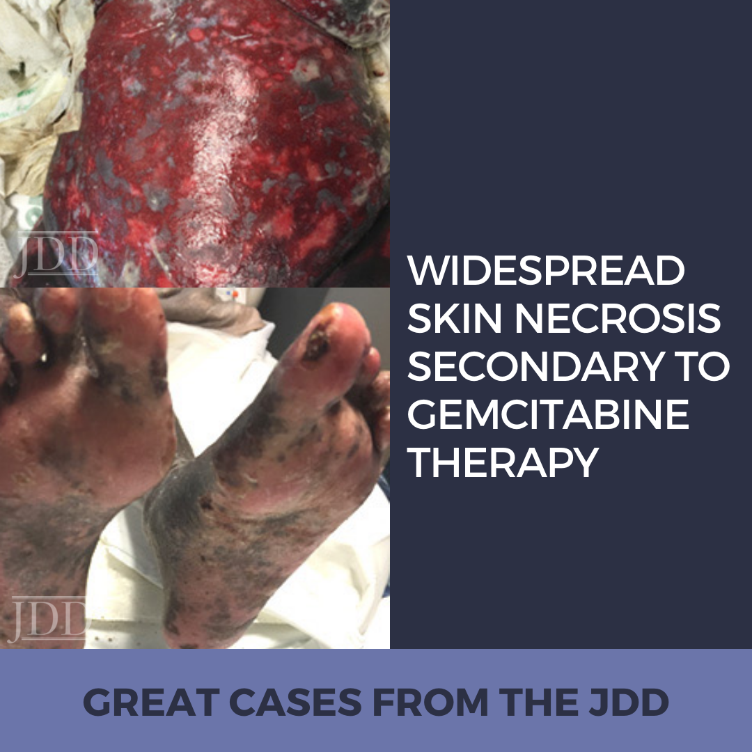
Patrick M. Zito DO PharmD, Adrianna M. Gonzalez BS, Joshua D. Fox MD, Megan Cronin MD, Nicholas Mackrides MD, Robert S. Kirsner MD PhD, and Anna J. Nichols MD PhD  (2018). Widespread Skin Necrosis Secondary to Gemcitabine Therapy 17(5).

https://jddonline.com/articles/dermatology/S1545961618P0582X

Content and images used with permission from the Journal of Drugs in Dermatology.

Adapted from original article for length and style.

Did you enjoy this case report? Find more here.

error

Enjoy this blog? Please spread the word :)