
Friday Pop Quiz 3/20/2026
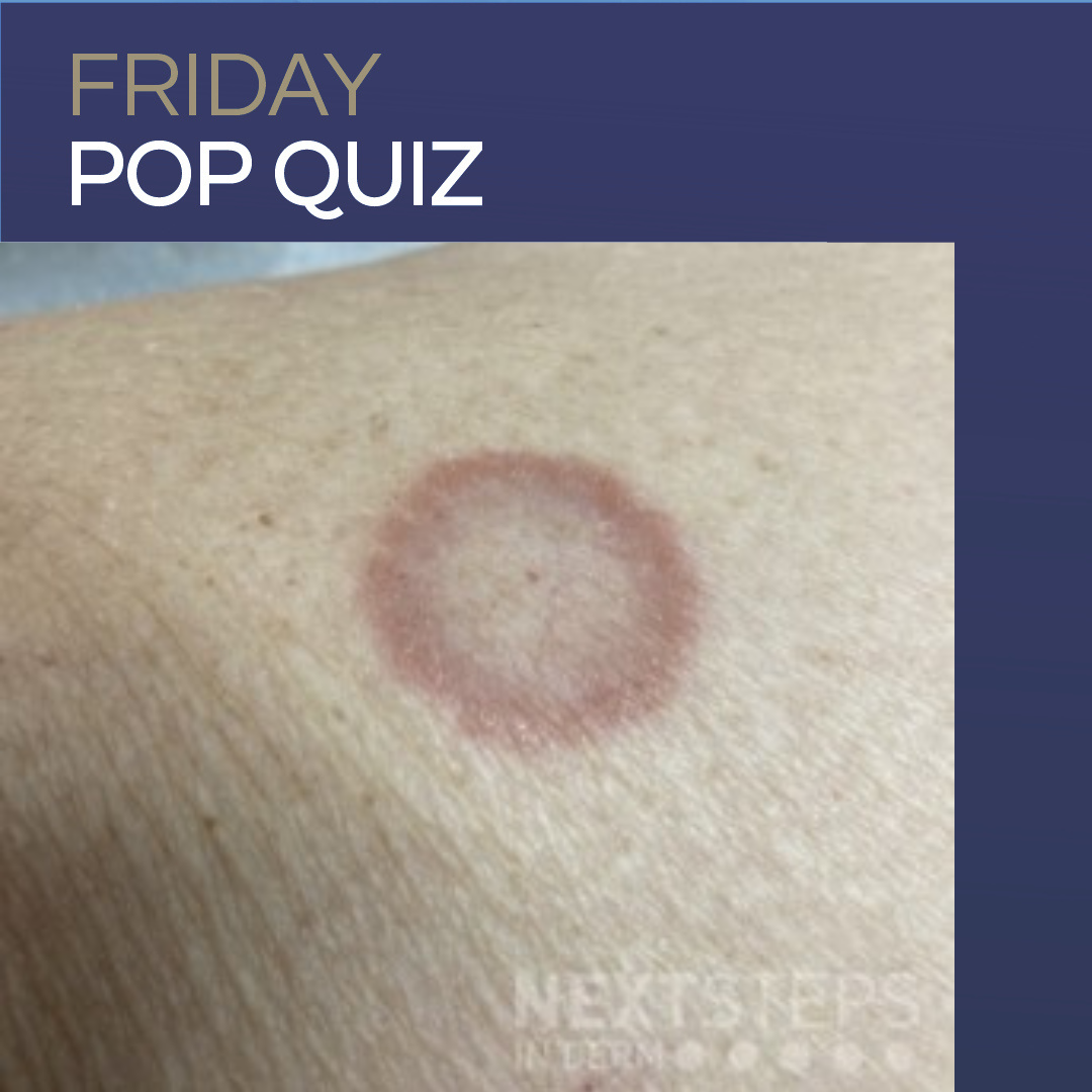 What histologic features are typically seen on a skin biopsy of this lesion?
A. Necrobiotic degeneration of dermal collagen surrounded by an inflammatory reaction
B. Layers with open-ended necrobiotic foci; lack of mucin; increased numbers of plasma cells (necrobiosis lipoidica)
C. Larger areas of eosinophilic necrobiosis and a lack of mucin deposition (rheumatoid nodule)
D. Immuno …
What histologic features are typically seen on a skin biopsy of this lesion?
A. Necrobiotic degeneration of dermal collagen surrounded by an inflammatory reaction
B. Layers with open-ended necrobiotic foci; lack of mucin; increased numbers of plasma cells (necrobiosis lipoidica)
C. Larger areas of eosinophilic necrobiosis and a lack of mucin deposition (rheumatoid nodule)
D. Immuno … 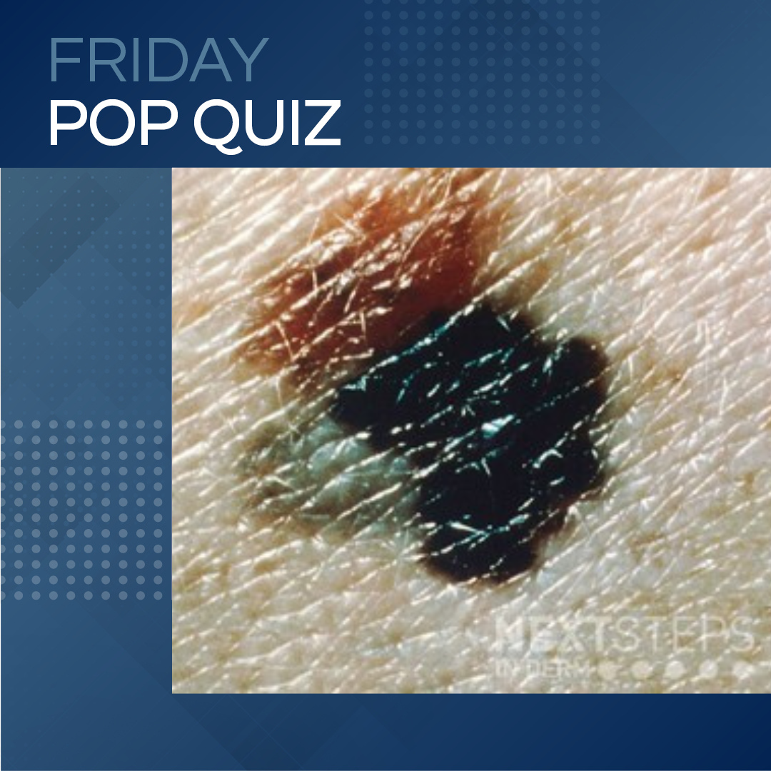 A patient presents with the lesion on the abdomen. The biopsy proves to be malignant melanoma with a breslow depth of 1.3mm. What is the tumor stage of this lesion?
A. T1a
B. T1b
C. T2a
D. T2b
E. T3a
To find out the correct answer and read the explanation, click here.
Brought to you by our brand partner
…
A patient presents with the lesion on the abdomen. The biopsy proves to be malignant melanoma with a breslow depth of 1.3mm. What is the tumor stage of this lesion?
A. T1a
B. T1b
C. T2a
D. T2b
E. T3a
To find out the correct answer and read the explanation, click here.
Brought to you by our brand partner
… 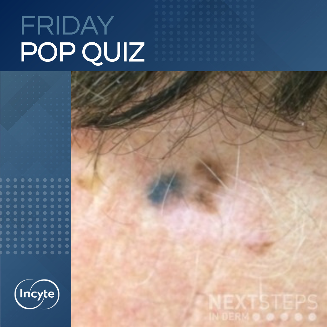 This patient presented for a different lesion but you see this during your exam and perform a biopsy. If the pigmented lesion seen here is 0.9mm deep, what is the recommended margin for excision?
A. <0.5cm
B. 0.5cm
C. 1.0cm
D. 2.0cm
E. >2.0cm
To find out the correct answer and read the explanation, click here.
Brought to you by our brand partner
…
This patient presented for a different lesion but you see this during your exam and perform a biopsy. If the pigmented lesion seen here is 0.9mm deep, what is the recommended margin for excision?
A. <0.5cm
B. 0.5cm
C. 1.0cm
D. 2.0cm
E. >2.0cm
To find out the correct answer and read the explanation, click here.
Brought to you by our brand partner
… 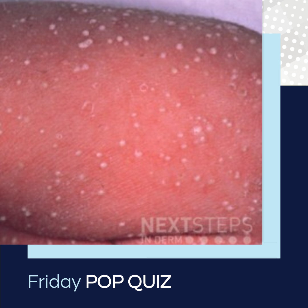 Which of the following cells is most likely to be seen in a biopsy of a lesion from this 1-day-old child?
A. Eosinophils
B. Histiocytes
C. Lymphocytes
D. Mast cells
E. Neutrophils
To find out the correct answer and read the explanation, click here. …
Which of the following cells is most likely to be seen in a biopsy of a lesion from this 1-day-old child?
A. Eosinophils
B. Histiocytes
C. Lymphocytes
D. Mast cells
E. Neutrophils
To find out the correct answer and read the explanation, click here. …  Next Steps in Derm, in partnership with ODAC Dermatology, Aesthetic and Surgical Conference, interviewed Dr. Vishal A. Patel (fellowship trained Mohs micrographic surgeon who serves as Director of Cutaneous Oncology at the GW Cancer Center and Director of Dermatologic Surgery at the GW Department of Dermatology) about routine skin cancer checks. Watch as he provides insight on this complex questio …