Grab a Fork and Knife: An Approach to Treating Rosacea
 For most dermatologic conditions, my mantra for treatment tends to be “less is more”. I prefer to use the fewest number of creams, pills, and steps to achieve the best results. However, after watching this year’s ODAC Sneak Peek Inflammatory Diseases Symposium, I may have a new mantra for treating my rosacea patients – “forks, spoons, and knives”.
At the ODAC 2021 Pre-Conference Sne …
 Continue reading "Grab a Fork and Knife: An Approach to Treating Rosacea"


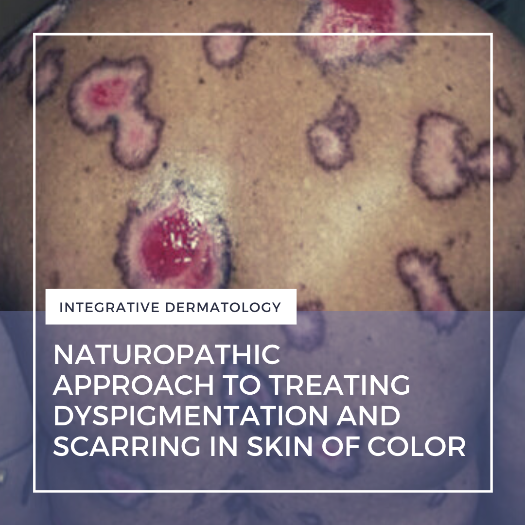
 As we know, people of color are more apt to have problems with dyspigmentation and excessive scarring (keloid formation and hypertrophic scars). We probably all use a combination approach for both of these: sun protection, peels, HQ, OTC skin lighteners, injections, lasers, energy-based devices, etc. These are common approaches, but what are the go-to’s for a naturopathic physician? You may ne …
  As part of the recent 2020 Integrative Dermatology Symposium, Dr. Jason Hawkes, Associate Clinical Professor of Dermatology at UC Davis, provided a fantastically comprehensive primer on the management of psoriasis in the COVID-19 era. Read on for important treatment recommendations and insights to improve your clinical practice!
As an introduction, Dr. Hawkes emphasized that psoriasis remains a …
  At the ODAC 2021 Sneak Peek Symposium on Inflammatory Skin Diseases, expert faculty presented on the topics of acne, rosacea, atopic dermatitis, hidradenitis suppurativa, and psoriasis. If you missed the live symposium, Next Steps will be sharing highlights and a summary of each lecture over the course of the next few weeks. Today, Dr. Blari Allais shares an excellent recap of Dr. Joslyn Kirby's s …
  CASE REPORT
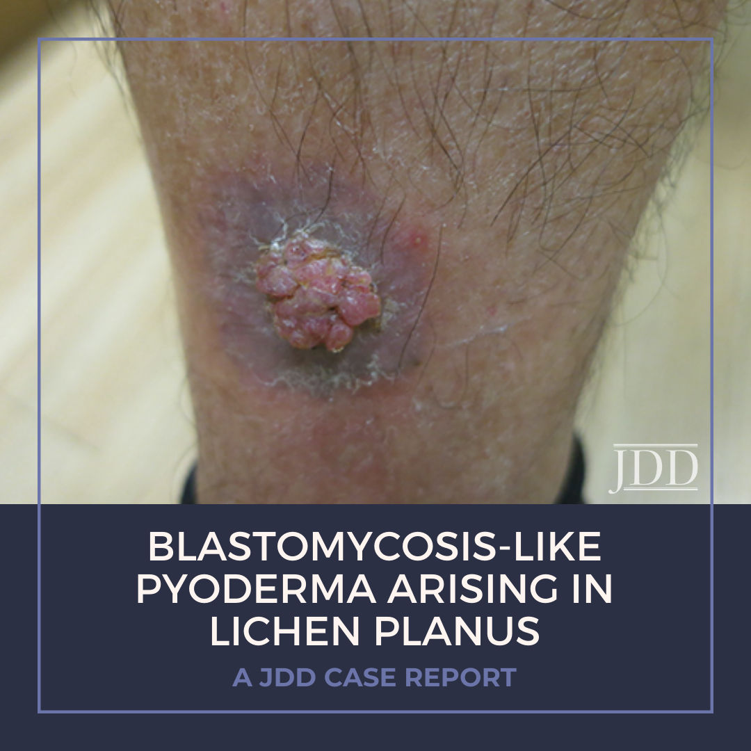
A 71-year-old Fitzpatrick phototype IV man with a history of hyperlipidemia and extensive travel to the Middle East presented with a mildly painful vegetative growth on his right lower leg for 1.5 months (Figure 1). In 2014, the patient reported a pruritic “rash” in the same location, which was treated with fluocinonide .05% ointment with resolution.
[caption id="attachment …