
Sam’s Severe Sweating Story | Hyperhidrosis Awareness Month
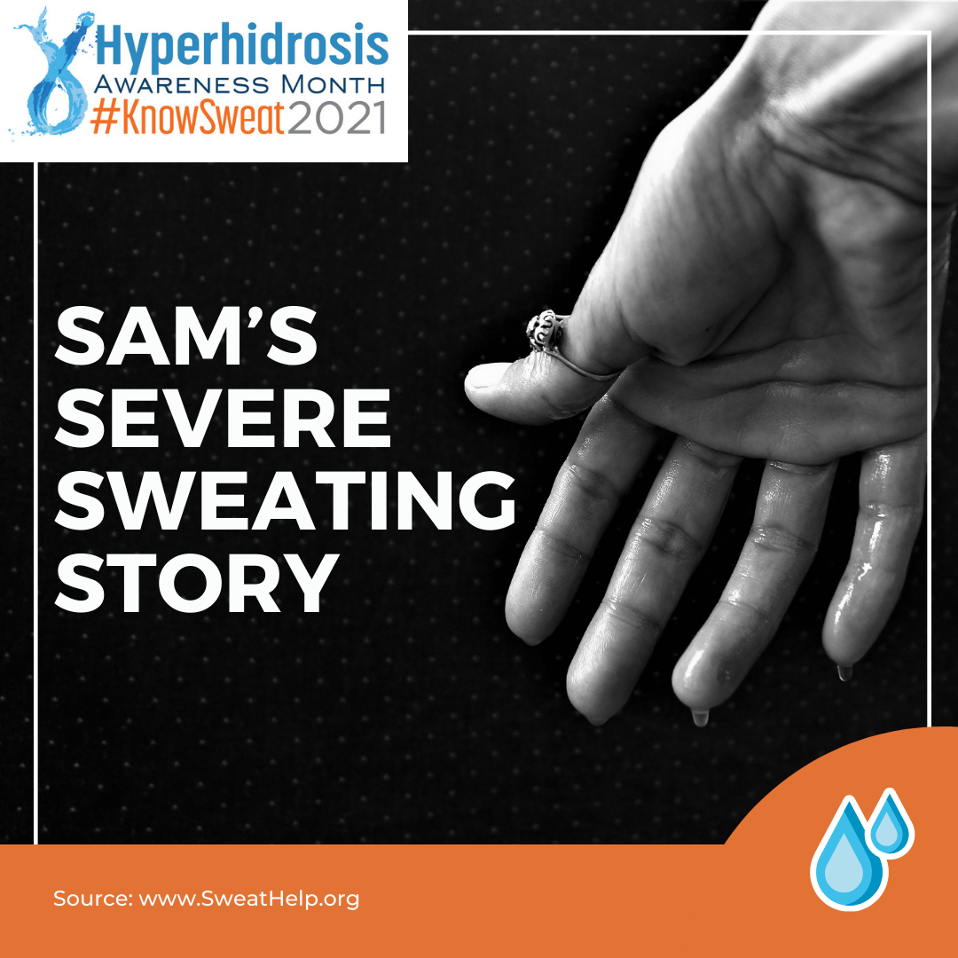 Sam’s Severe Sweating Story
Like many people with primary hyperhidrosis (Hh) or excessive, uncontrollable sweating, I am most affected by Hh on the palms of my hands, soles of my feet, and in my underarms. My chronic extreme sweating isn’t caused by an underlying medical condition and it can occur at any time, even when I’m not hot, exercising, or in a stressful situation. Without treatme …
Sam’s Severe Sweating Story
Like many people with primary hyperhidrosis (Hh) or excessive, uncontrollable sweating, I am most affected by Hh on the palms of my hands, soles of my feet, and in my underarms. My chronic extreme sweating isn’t caused by an underlying medical condition and it can occur at any time, even when I’m not hot, exercising, or in a stressful situation. Without treatme … Continue reading "Sam’s Severe Sweating Story | Hyperhidrosis Awareness Month"
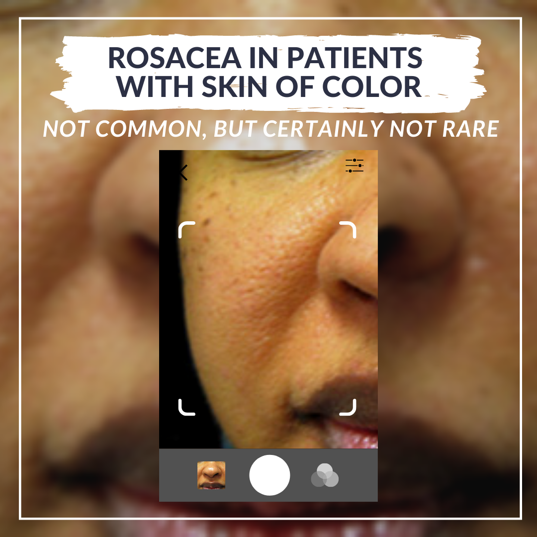 Rosacea is defined by facial erythema and telangiectasias, two features that are more difficult to appreciate in patients with skin of color. To refine our ability to diagnose rosacea in all skin types, on day two of the 2021 Skin of Color Update virtual conference, we had an informative, evidence-based lecture, “Recognizing, Diagnosing, and Treating Rosacea in Patients with Skin of Color,” by …
 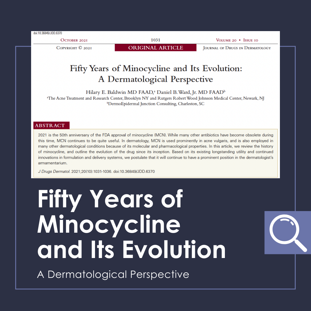 2021 is the 50th anniversary of the FDA approval of minocycline (MCN). While many other antibiotics have become obsolete during this time, MCN continues to be quite useful. In dermatology, MCN is used prominently in acne vulgaris, and is also employed in many other dermatological conditions because of its molecular and pharmacological properties. In this article, we review the history of minocycli …
 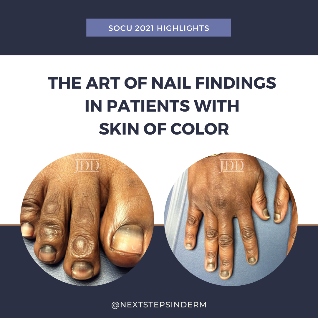 Dr. Shari Lipner, Associate Professor of Clinical Dermatology and Director of the Nail Division at Weill Cornell Medicine and President of The Dermatologic Society of Greater New York, shared her expertise of nail disorders in patients with skin of color: from nail psoriasis and onychomycosis to subungual melanoma.
Dr. Lipner’s lecture focused on the following key points (spoiler alert!):
…
Dr. Shari Lipner, Associate Professor of Clinical Dermatology and Director of the Nail Division at Weill Cornell Medicine and President of The Dermatologic Society of Greater New York, shared her expertise of nail disorders in patients with skin of color: from nail psoriasis and onychomycosis to subungual melanoma.
Dr. Lipner’s lecture focused on the following key points (spoiler alert!):
…  As dermatologists, we understand that hidradenitis suppurativa (HS) can be a devastating disease that affects all aspects of our patients’ lives, influencing their physical health, ability to be active, and ability to be intimate. This disease disproportionately affects patients with skin of color and unfortunately, treatment can be challenging. We are lucky to have experts who dedicate their cl …