
Lamotrigine – Friday Pop Quiz 3/6
 This 20-year-old woman started lamotrigine 3 weeks ago. She takes no other medications, and has no comorbid conditions. Her urine b-HCG is positive. Which of the following treatments has the theoretically lowest fetal risk?
A. Prednisone
B. IVIG
C. Etanercept
D. Cyclosporine
E. Rituximab
To find out the correct answer and read the explanation, click here.
Brought to you by our …
This 20-year-old woman started lamotrigine 3 weeks ago. She takes no other medications, and has no comorbid conditions. Her urine b-HCG is positive. Which of the following treatments has the theoretically lowest fetal risk?
A. Prednisone
B. IVIG
C. Etanercept
D. Cyclosporine
E. Rituximab
To find out the correct answer and read the explanation, click here.
Brought to you by our … 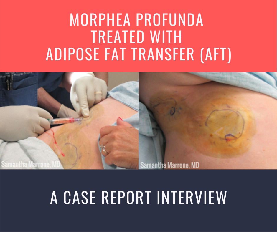 Morphea profunda. To the dermatologist, these words conjure images of hyperpigmented to violaceous, indurated, bound down atrophic plaques. We describe these lesions in our specialty’s vernacular, so that those we are conversing with can often surmise the diagnosis before even seeing the affected patient. But to the patient, it is the language of the diagnosis itself that has the most meaning. M …
 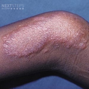 What would this lesion show histologically?
A. A proliferation of basaloid cells with small ducts
B. A grenz zone with a lot of inflammatory cells and globi
C. Well formed tuberculoid granulomas in a linear pattern
D. Scattered comedo-like cysts
E. Acanthosis and neutrophilic infiltrate
To find out the correct answer and read the explanation, click here.
Brought to you by our …
What would this lesion show histologically?
A. A proliferation of basaloid cells with small ducts
B. A grenz zone with a lot of inflammatory cells and globi
C. Well formed tuberculoid granulomas in a linear pattern
D. Scattered comedo-like cysts
E. Acanthosis and neutrophilic infiltrate
To find out the correct answer and read the explanation, click here.
Brought to you by our …  Well+Good recently published an article asking if hair loss supplements actually work.
For an expert opinion, I consulted Crystal Aguh, MD, Director of the Ethnic Skin Program and Assistant Professor in the Department of Dermatology at Johns Hopkins University School of Medicine, Baltimore, MD.
What is the theory behind hair supplements?
Hair supplements are designed to create an ideal nu …
Well+Good recently published an article asking if hair loss supplements actually work.
For an expert opinion, I consulted Crystal Aguh, MD, Director of the Ethnic Skin Program and Assistant Professor in the Department of Dermatology at Johns Hopkins University School of Medicine, Baltimore, MD.
What is the theory behind hair supplements?
Hair supplements are designed to create an ideal nu …  Next Steps in Derm, in partnership with ODAC Dermatology, Aesthetic and Surgical Conference, is excited to share a new video series where dermatology key opinion leaders share important updates and pearls on a variety of medical, surgical, and aesthetic dermatology topics.
First up is Dr. Amy McMichael, Professor and Chair of Dermatology at the Wake Forest University School of Medicine, sharing …
Next Steps in Derm, in partnership with ODAC Dermatology, Aesthetic and Surgical Conference, is excited to share a new video series where dermatology key opinion leaders share important updates and pearls on a variety of medical, surgical, and aesthetic dermatology topics.
First up is Dr. Amy McMichael, Professor and Chair of Dermatology at the Wake Forest University School of Medicine, sharing …