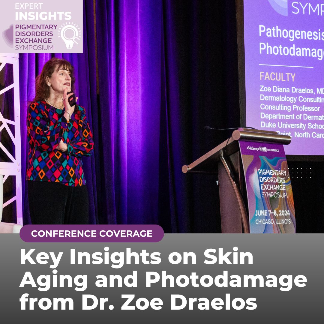
At the 2024 Pigmentary Disorders Exchange Symposium in Chicago, Dr. Zoe Draelos, an expert dermatologist and research scientist, gave a comprehensive lecture on skin aging, emphasizing that photodamage is just one aspect of a broader spectrum of factors influencing skin health. Her lecture focused on understanding how various elements contribute to skin aging beyond traditional UV radiation, including the role of pigmentation and other environmental factors.
When thinking about skin aging, most people consider extrinsic factors, such as UV radiation and smoking, and intrinsic factors, such as stress. However, Dr. Draelos introduces a new concept: the exposome. To better understand aging, we should now consider three dimensions of the exposome: the extrinsic exposome, the intrinsic exposome, and the epigenetic exposome.
The Exposome and Skin Aging
The exposome encompasses all environmental and biologic factors impacting skin aging throughout a person’s life. Damage to the exposome from various exposure events accumulates over the lifetime, and it even modifies our gut and skin microbiome making it less diverse, further contributing to aging.
The extrinsic exposome includes external factors such as UV radiation and pollution that contributes to extrinsic aging; the intrinsic exposome includes internal processes such as genetic changes and biochemical disruptions contributing to aging; and the third component is the environmental and biologic interplay, that is epigenetics and how our genome, epigenome and transcriptome influence aging.
Seven Exposome Factors Contributing to Skin Aging
EXTRINSIC EXPOSOME
-
- Photodamage
- UV radiation is a well-known cause of skin damage, but this is only one part of the story.
- Pollution
- Particulate matter and heavy metals can accelerate the aging process by inducing inflammation and oxidative stress within our skin
- Smoking/Tobacco
- Combustion byproducts, like nanoparticles from pollution and tobacco smoke, generate oxidative stress on the skin and deplete the skin’s natural antioxidants, accelerating aging.
- Heat
- High temperatures can also exacerbate skin aging by disrupting the skin barrier and promoting break down of collagen.
- Photodamage
INTRINSIC EXPOSOME
-
- Poor Diet/Nutrition
- Adequate nutrition is vital for skin repair and maintenance.
- Stress
- Chronic stress disrupts the Hypothalamic-Pituitary Axis (HPA) and triggers release stress hormones that degrade the skin’s collagen and elastin, contributing to aging.
- Lack of Sleep
- HPA Irregularities can also be caused by insufficient sleep, and contribute to aging by interfering with the skin’s natural repair processes.
- Poor Diet/Nutrition
BIOLOGIC EXPOSOME
-
- This represents the cumulative effect of the interplay of all seven factors on our gene expression. This highlights how lifestyle, environmental exposures, and even psychologic stressors can influence the way genes are turned off and on, contributing to visible signs of aging, which stems from a biomolecular level.
What do These Seven Factors Have in Common?
They all produce Reactive Oxygen Species (ROS), which are reactive molecules containing oxygen that can cause damage to cells, proteins, lipids, and DNA.
Aging results from chronic production of ROS, which leads to chronic inflammation, skin damage and a negative effect on our exposome.
How to Combat Reactive Oxygen Species?
Dr. Draelos shared encouraging news that we can mitigate the damage caused by exposomes through the use of adaptogens.
Adaptogens: “A non-toxic substance and especially a plant extract that is held to increase the body’s ability to resist the damaging effects of stress and promote or restore normal physiological functioning” –Merriam-Webster
She explained that adaptogens are essentially a new term for antioxidants that help protect the exposome by neutralizing ROS. Examples include ashwagandha, green tea, and substances containing flavanols and polyphenols.
Biochemical Changes Affecting Aging
Dr. Draelos discussed biochemical processes that are disrupted with aging in the exposome. She went over these elements and defined them as follows:
-
- Genome: The complete set of genetic instructions, also known as the DNA, that builds and maintains our body (i.e., pigment, height). UV radiation can damage the genome, leading to skin cancer if replication is impaired.
- Epigenome: This consists of chemical modifications to the DNA and histone protein that regulate gene expression in different cells and tissues, influencing how the genome is used without changing the genetic sequence. Epigenetic changes are dynamic and damage here can activate genes at inappropriate times, impacting skin health.
- Transcriptome: The complete collection of RNA transcripts produced by the genome at any given time, representing the genes that are actively expressed. Changes can affect protein synthesis and skin integrity.
- Proteome: The entire set of functional proteins produced by our bodies. The proteome determines which collagen proteins are produced in the skin and reflects overall skin health.
- Metabolome: Includes all the chemicals or metabolites in our body or within a specific tissue, such as the skin, at any given time. This includes all the raw material, such as amino acids and vitamins, necessary for protein synthesis, energy production, and overall metabolic health. It is important for skin health maintenance and is influenced by diet, disease, stress, and environmental exposures.
Fitzpatrick Skin Type Affects the Skin’s Ability to Defend Against Damage
Understanding the exposome’s impact on skin aging is crucial. The skin is the first line of defense against damage to the exposome and aging, but the level of protection it offers varies across different Fitzpatrick skin types (FST). Differences in the composition of the stratum corneum, skin lipids, skin pH, the dermal structure, and melanin type and distribution among FSTs contribute to varying susceptibilities to aging and environmental damage. These differences dictate how effectively each skin type is able to resist the effects of aging.
Dr. Draelos defines African American skin as FST IV-VI, Hispanic skin as FST III, and Caucasian skin as FST I-II.
The first layer of exposome protection is the outermost layer of the skin—the stratum corneum.
-
- It is denser in African American skin, which results in less UV penetration, enhanced light scattering, and subsequently less ROS and dermal collagen damage compared to Caucasian skin tones.
- This layer also turns over 2.5 times faster in African American skin than Caucasian skin, providing further protection against UV radiation.
African American skin has a higher lipid content compared to Caucasian skin resulting in better moisturization. Interestingly, ceramide concentration is 50% lower in African American skin compared to Hispanic and Caucasian skin, which weakens the intracellular lipids that keep corneocytes together. This is why ceramide-containing moisturizers play a role in maintaining an optimal skin barrier.
Protecting the dermis from photodamage is extremely important for aging. The dermis contains most of the blood supply, which is where ROS are present.
-
- In African American skin, the dermal-epidermal junction is thicker corresponding to more tightly packed collagen and better protection against UV damage and therefore less photoaging.
- In contrast, Caucasians have a thinner dermal-epidermal junction, allowing deeper UV penetration and more photoaging.
The exposome is also protected by melanin, which has two forms in different concentrations depending on skin type:
-
- Eumelanin predominates in darker skin types (FST IV-VI) and provides effective UV protection by donating its extra electron to UV-induced ROS and thereby neutralizing it, acting as an antioxidant. In doing so, the melanin is oxidized and a tan results.
- Pheomelanin is more common in lighter skin types (FST I-II) and lacks eumelanin’s protective ability because it does not have an extra electron available to neutralize ROS. Therefore, it is actually a prooxidant that increases ROS with photo exposure leading to a higher susceptibility to sunburn, skin damage, and eventually skin cancer.
The distribution of melanin and the location of the UV protective layer differs between skin types affecting how the skin ages and protects itself against UV damage. The deeper the protective layer within the skin, the better it protects the dermis against UV damage.
-
- African American skin has more melanin in the basal layer and has a UV filter that is deep in the basal and spinous layers, providing strong protection.
- Caucasian skin has melanin closer to the surface, making it more susceptible to damage if that layer is removed. The UV filter is mostly in the stratum corneum.
- Because of this, exfoliation methods like using glycolic acid or microdermabrasion, which remove this protective layer in Caucasian skin, making the skin more vulnerable to UV damage, necessitating the use of sunscreen.
Additionally, the number of melanosomes and their pH differ between African American skin and Caucasian skin.
-
- African American skin contains a greater number of melanosomes (~200) within the melanocytes. These melanosomes are pH neutral.
- Caucasian skin contains less melanosomes (~20), which are more acidic.
The pH matters because tyrosinase activity is pH dependent with neutral pHs leading to greater pigment production.
Implications for Sunscreen Use in Combatting Aging
Without sunscreen, the natural protection against UV-A and UV-B rays differs between African American and Caucasian skin due to the inherent physiologic differences that were just reviewed.
-
- African American skin has an endogenous UVA protection of 5.7, whereas in Caucasians, it is 1.8 meaning that 4x more UVA reaches the upper dermis of Caucasian compared to African American skin.
- African American skin has an endogenous UVB protection level, or SPF, of 13.4 versus SPF 3.4 in Caucasian skin.
Dr. Draelos notes that companies are taking efforts to enhance sunscreen use in individuals with darker skin tones by grinding zinc oxide and titanium dioxide, which normally leave a white residue, into smaller transparent forms. However, this reduces their UV reflection and lowers SPF.
Iron oxide is newer ingredient that has been added into sunscreens due to this pigment’s ability to absorb and reflect visible light, preventing pigment darkening in darker complexions. Black, red, and yellow iron oxides can be used to tint sunscreens to match various skin tones while maintaining UV protection.
Sunscreen helps protect against photoinduced skin damage, but it is also effective against environmental pollutants. Sunscreens and moisturizers can protect against combustion nanoparticles by forming a film over the skin that prevents them from penetrating the skin and causing damage.
Lifestyle
To conclude the discussion, Dr. Draelos reminded the audience of the importance of sleep, stress management, and good nutrition in maintaining a healthy immune system and microbiome. A weakened immune system and disrupted microbiome accelerate aging. Choosing vegetables high in Vitamin C can help replenish the skin’s antioxidant activity and neutralize ROS.
In summary, Dr. Draelos emphasized that understanding skin aging requires a comprehensive approach. The exposome model highlights the cumulative effects of various factors on skin health. Adaptogens and modern sunscreen formulations play crucial roles in protecting against skin aging. By considering pigmentation and the diverse needs of different skin types, dermatologists can better address skin aging and maintain overall skin health.
REFERENCES:
-
- Krutmann J, Bouloc A, Sore G, Bernard BA, Passeron T. The skin aging exposome. J Dermatol Sci. 2017;85(3):152-161. doi:10.1016/j.jdermsci.2016.09.015.
- Jin SP, Li Z, Choi EK, et al. Urban particulate matter in air pollution penetrates into the barrier-disrupted skin and produces ROS-dependent cutaneous inflammatory response in vivo. J Dermatol Sci. 2018;91:175-183.
- Kaidbey KH, Agin PP, Sayre RM, Kligman AM. Photoprotection by melanin–a comparison of black and Caucasian skin. J Am Acad Dermatol. 1979;1:249-260.
- Weigand DA, Haygood C, Gaylor JR. Cell layers and density of Negro and Caucasian stratum corneum. J Investig Dermatol. 1974;62(6):563-568. doi:10.1111/1523-1747.ep12679412.
- Corcuff P, Lotte C, Rougier A, Maibach HI. Racial differences in corneocytes. A comparison between black, white, and oriental skin. Acta Derm Venereol. 1991;71(2):146-148. doi:10.2340/0001555571146148.
- Muizzuddin N, Hellemans L, Van Overloop L, Corstjens H, Declercq L, Maes D. Structural and functional differences in barrier properties of African American, Caucasian, and East Asian skin. J Dermatol Sci. 2010;59(2):123-128. doi:10.1016/j.jdermsci.2010.06.003.
- Md Jaffri J. Reactive oxygen species and antioxidant system in selected skin disorders. Malaysian J Med Sci. 2023;30(1):7-20. doi:10.21315/mjms2023.30.1.2.
- Girardeau S, Mine S, Pageon H, Asselineau D. The Caucasian and African skin types differ morphologically and functionally in their dermal component. Exp Dermatol. 2009;18(8):704-711. doi:10.1111/j.1600-0625.2009.00843.x.
- Querleux B, Baldeweck T, Diridollou S, et al. Skin from various ethnic origins and aging: an in vivo cross-sectional multimodality imaging study. Skin Res Technol. 2009;15(3):306-313. doi:10.1111/j.1600-0846.2009.00365.x.
- Del Bino S, Ito S, Sok J, et al. Chemical analysis of constitutive pigmentation of human epidermis reveals constant eumelanin to pheomelanin ratio. Pigment Cell Melanoma Res. 2015;28(6):707-717. doi:10.1111/pcmr.12410.
- Thong HY, Jee SH, Sun CC, Boissy RE. The patterns of melanosome distribution in keratinocytes of human skin as one determining factor of skin colour. Br J Dermatol. 2003;149(3):498-505. doi:10.1046/j.1365-2133.2003.05473.x.
- Fuller BB, Spaulding DT, Smith DR. Regulation of the catalytic activity of preexisting tyrosinase in black and Caucasian human melanocyte cell cultures. Exp Cell Res. 2001;262(2):197-208. doi:10.1006/excr.2000.5092.
- Seck S, Hamad J, Schalka S, Lim HW. Photoprotection in skin of color. Photochem Photobiol Sci. 2023;22(2):441-456. doi:10.1007/s43630-022-00314-z.
- Bernstein EF, Sarkas HW, Boland P, Bouche D. Beyond sun protection factor: An approach to environmental protection with novel mineral coatings in a vehicle containing a blend of skincare ingredients. J Cosmet Dermatol. 2020;19(2):407-415. doi:10.1111/jocd.13007.
- Puri P, Nandar SK, Kathuria S, Ramesh I. Effects of air pollution on the skin: a review. Indian J Dermatol Venereol Leprol. 2017;83:415-423.
- Krutmann J, Liu W, Li L, et al. Pollution and skin: from epidemiological and mechanistic studies to clinical implications. J Dermatol Sci. 2014;76:163-168.
This information was presented by Dr. Zoe Draelos during the 2024 Pigmentary Disorders Exchange Symposium. The above session highlights were written and compiled by Dr. Sarah Millan.
Did you enjoy this article? You can find more here.

