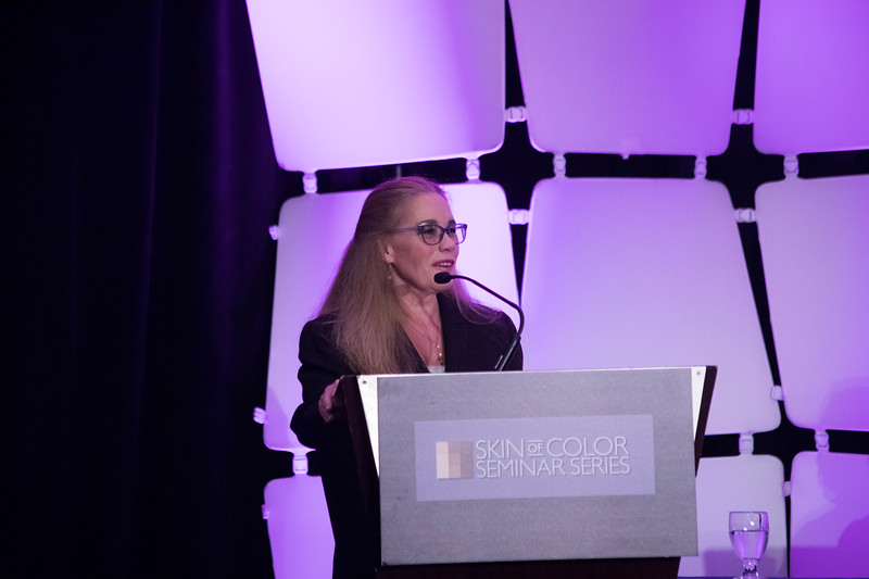
This article features a recap of Dr. Hilary Baldwin’s talk on the etiology, risk factors, and treatment of keloids at the 2018 Skin of Color Seminar Series, now known as the Skin of Color Update. Dr. Bridget Kaufman, onsite correspondent for the meeting, shares highlights directly from the talk. Dr. Baldwin focused on earlobe keloid scars in particular, which may present with several different morphologies: anterior button, posterior button, wraparound, dumbbell, and lobular.
Dr. Baldwin started by discussing why some patients are prone to keloids and others do not. Based on a study of 220 patients at Kings County Hospital, there appears to be no difference in rate of cartilage piercing, metal sensitivity, piercing sites, types of earrings worn, piercing method, hormonal influences, or age at piercing between keloid formers and non-keloid formers. In the keloid former group, 12.8% of patients developed keloids at the first piercing, and the risk of keloid formation dramatically increased at each piercing thereafter (70.2% risk for 2nd piercing).
Important Clinical Pearls:
-
Ear pierces on babies have a 0% risk of keloids
Piercings done pre-menarche have a significantly lower risk of keloids than those performed post-menarche
First pierces rarely keloid
The chances of subsequent pierce keloiding in a keloid former is at least 20% or higher
Dr. Baldwin then moved on to the treatment of keloids. Earlobe keloids are significantly easier to treat than classic keloids on the body. This is because earlobes are a discrete tissue with little to no tension, pressure dressings can be used, and patients tend to be very motivated and compliant.
For surgery alone, recurrence rate on the ear is 39-42% (vs. 100% on body). Surgery plus corticosteroids and surgery with radiation therapy for earlobe keloids are associated with a 1-3% (vs. 50% on body) and 0-25% (same for body) risk of recurrence.
Imiquimod helps prevent keloid recurrence when used on the earlobes, but does not appear to be effective on other body parts. It can be used as part of the healing process.
Dr. Baldwin has developed her own method for excising dumbbell keloids called dumbbell keloids for dummies:
- Shave off anterior button
- Shave off posterior button
- Measure diameter of keloid core
- Select punch to be at least 1mm wider than core
- Stabilize and punch through to a tongue depressor
- Suture right/left anteriorly and superior/inferior posteriorly
Unfortunately, not all keloids can be surgically excised. The major dogma of keloid surgery is “don’t leave any keloid tissue behind.” Hilary’s dogma of earlobe keloid surgery is “a non-functional earlobe is a treatment failure. Therefore, Dr. Baldwin has looked towards alternative treatments for keloids in such cases. She performed a study in 5 patients in whom complete removal of keloids would leave a non-functional earlobe. She sculpted the keloid to earlobe shape, injected interferon-alpha 2b 1.5-million units/cm, covered the wound with a compression earring, and allowed for secondary intention healing. In the patients who completed therapy, there was no uncontrollable recurrence at 4-6 year follow-up, although all patients are continuing the use of pressure earrings.
The combination of targeted keloid removal, interferon-alpha 2b injection, and pressure earrings may be an option for patients with large, difficult-to-treat keloids.
For those with a history of keloids, re-piercing and tattoos should be performed with caution. As Dr. Baldwin mentioned previously, there is a 20% chance of re-keloiding with subsequent piercings; however, this risk can be somewhat reduced with corticosteroid and interferon injections.
Keloids do occur at tattoo sites, but it is very uncommon.
Most tattoo keloids have been reported in patients who perform ritual keloiding in Africa. Nonetheless, trauma from tattoos, repeated passages, and placement in high risk sites or high risk patients may result in keloid formation!
For tattoo removal, Nd:YAG is the best laser therapy and appears to be associated with a very low (if no) risk of keloid formation.
This was a very informative lecture on earlobe keloids. I now feel more equipped to counsel my patients on the risk factors for and treatment of earlobe keloids. I hope that you do too!


