
Ocular Dermoid in Patient With Basal Cell Nevus Syndrome
 CASE
A 47-year-old woman presented for Mohs Micrographic Surgery for a biopsy-proven basal cell carcinoma involving the right nasal ala. The patient had a history of basal cell nevus syndrome (BCNS) and previous history of multiple basal cell carcinomas.
On initial examination, the patient was noted to have a few scattered pearly molluscoid papules on the head and neck, which were suspicio …
CASE
A 47-year-old woman presented for Mohs Micrographic Surgery for a biopsy-proven basal cell carcinoma involving the right nasal ala. The patient had a history of basal cell nevus syndrome (BCNS) and previous history of multiple basal cell carcinomas.
On initial examination, the patient was noted to have a few scattered pearly molluscoid papules on the head and neck, which were suspicio …
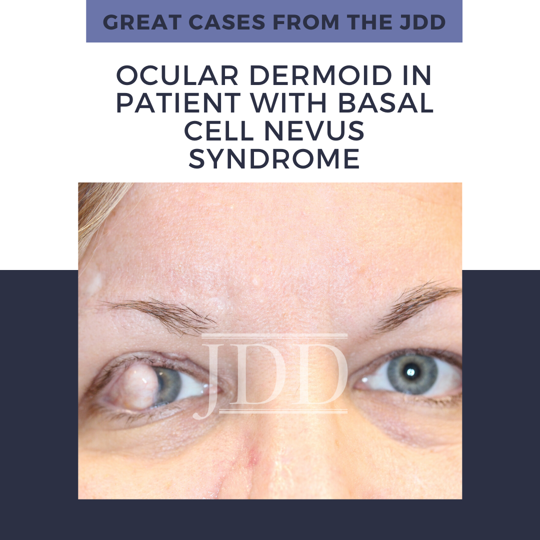
CASE
A 47-year-old woman presented for Mohs Micrographic Surgery for a biopsy-proven basal cell carcinoma involving the right nasal ala. The patient had a history of basal cell nevus syndrome (BCNS) and previous history of multiple basal cell carcinomas.
On initial examination, the patient was noted to have a few scattered pearly molluscoid papules on the head and neck, which were suspicio … Continue reading "Ocular Dermoid in Patient With Basal Cell Nevus Syndrome"
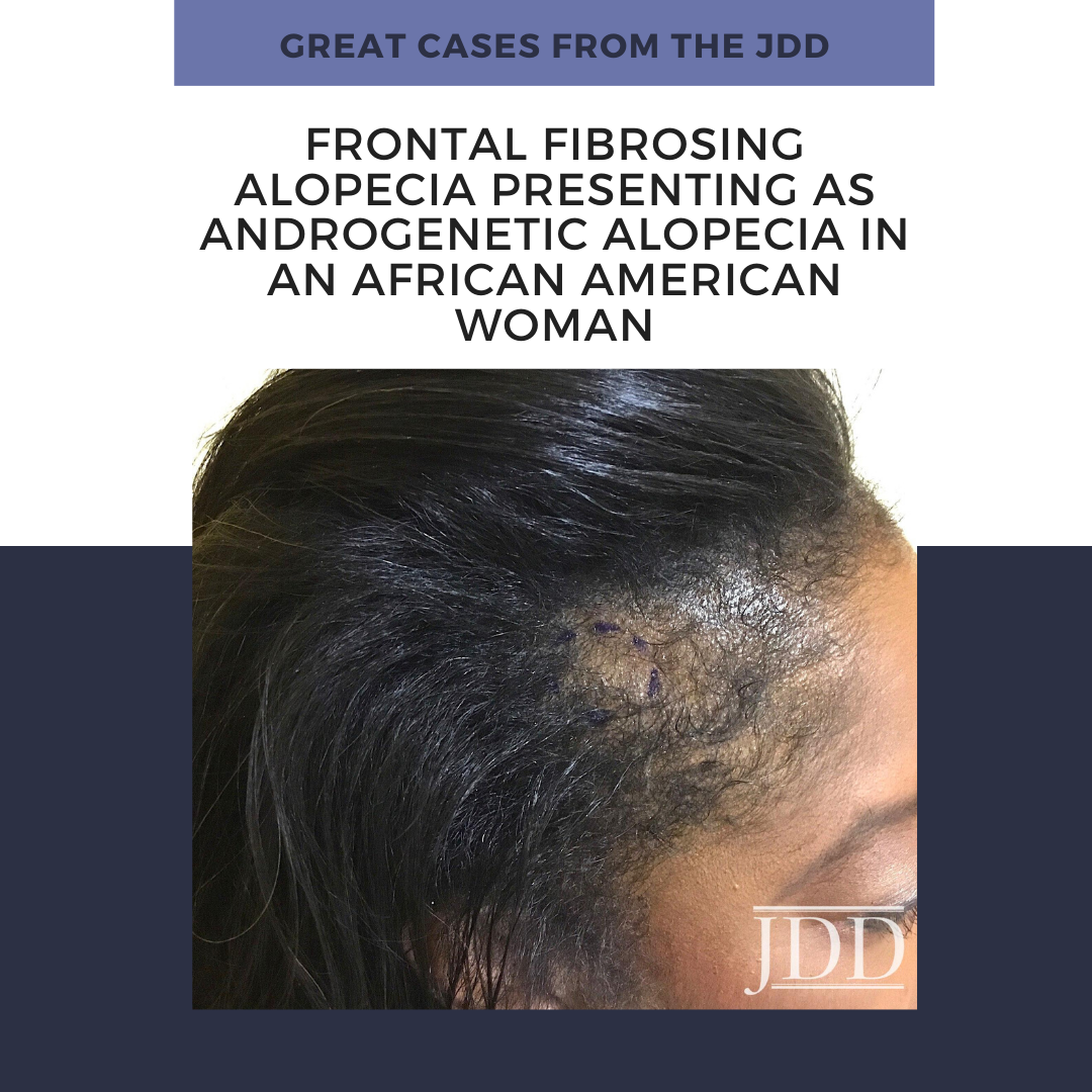 Frontal fibrosing alopecia (FFA) is a primary lymphocytic cicatricial alopecia that is currently regarded as a variant of lichen planopilaris. FFA has historically been considered rare in black patients, in whom traction alopecia, central centrifugal cicatricial alopecia, and androgenetic alopecia are frequently assumed to be more common. JDD author Kimberly Huerth, MD, ME describes a case of FFA …
 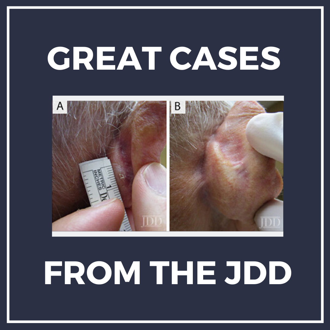 JDD Authors Rachel Fayne BA, Sonali Nanda MS, Anna Nichols MD PhD, and John Shen MD report a case of biopsy-proven invasive SCC in an 86-year-old Caucasian male with history of multiple actinic keratoses and no previous skin cancers. The patient declined surgical treatment due to concerns about cosmetic outcomes. A combination of topical 5% imiquimod cream, 2% 5-FU solution, and 0.1% tretinoin cre …
 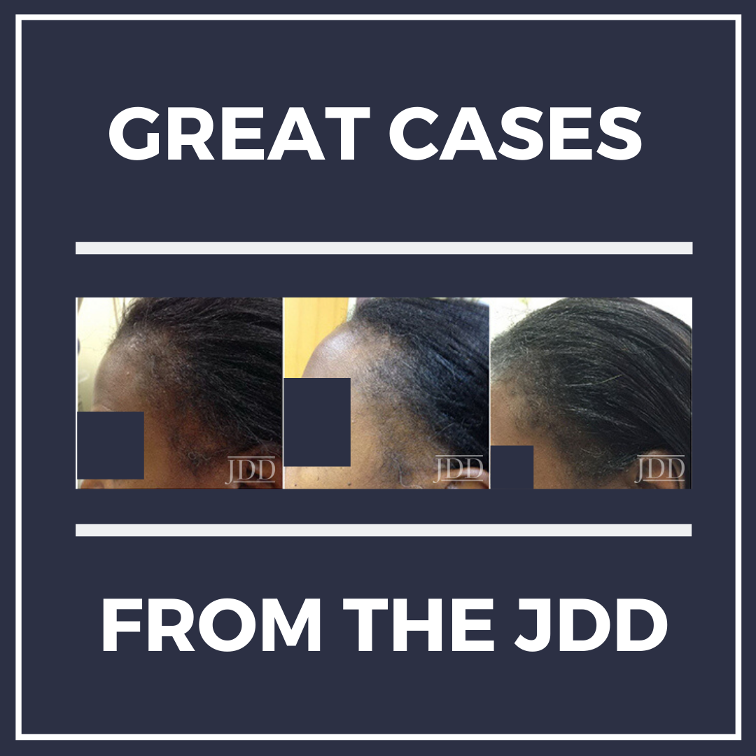 In this case series, JDD authors evaluate the efficacy and safety of intralesional triamcinolone acetonide injections (ILK) when used with topical minoxidil in the management of traction alopecia in 6 African American women.
Background
Traction alopecia (TA) is a form of hair loss secondary to repetitive and/or prolonged tension to a hair follicle over an extended period of time. This typically …
In this case series, JDD authors evaluate the efficacy and safety of intralesional triamcinolone acetonide injections (ILK) when used with topical minoxidil in the management of traction alopecia in 6 African American women.
Background
Traction alopecia (TA) is a form of hair loss secondary to repetitive and/or prolonged tension to a hair follicle over an extended period of time. This typically … 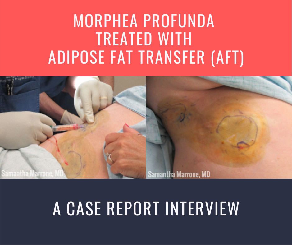 Morphea profunda. To the dermatologist, these words conjure images of hyperpigmented to violaceous, indurated, bound down atrophic plaques. We describe these lesions in our specialty’s vernacular, so that those we are conversing with can often surmise the diagnosis before even seeing the affected patient. But to the patient, it is the language of the diagnosis itself that has the most meaning. M …