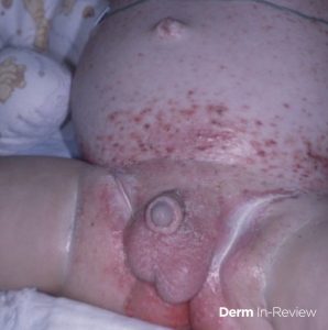
A 24 month-old infant presents with red-brown, crusted papules with petechiae in a seborrheic distribution. A biopsy is done to confirm a diagnosis. Which histologic picture is most likely?
A. CD1-, S100- cells with reniform nuclei
B. Foamy histiocytes with Touton giant cells
C. CD1+, S100+ cells with reniform nuclei
D. Mixed cellular infiltrate in a “ball and claw” pattern
E. Superficial perivascular infiltrate with mild spongiosis and neutrophil containing scale crust
To find out the correct answer and read the explanation, click here.
Brought to you by our brand partner Derm In-Review. A product of SanovaWorks.
![]()
