

Dr. Susan Weinkle, an expert in the field of aesthetic and surgical dermatology, shares important anatomical concepts to consider when using neurotoxins and fillers safely.
The face can be split into 3 zones – the upper 1/3rd, the middle 1/3rd and the lower 1/3rd. Knowing the important vessels and nerves in each zone as well as their corresponding depth in the skin is crucial in order to minimize injury and utilize a safe technique when injecting neurotoxins and fillers.
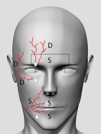
Figure 1: D corresponds to areas where deeper injections are safer to avoid vasculature and nerves. S corresponds to areas where superficial (intradermal injections) are safer to avoid vasculature and nerves. Vessels shown are cartoon depictions of some of the more common arteries including the supraorbital, supratrochlear, superficial temporal, facial and superior and inferior labial arteries. Also depicted are the supraorbital, infraorbital and mental foramen that are located along the medial limbus.
Important Landmarks for the Upper 1/3rd of the Face
- A line drawn vertically along the medial limbus denotes the anatomical locations of the supraorbital, infraorbital and mental foramen.
- The supraorbital notch or foramen is the exit aperture for the supraorbital neurovascular bundle (NVB). While the vessels here initially lie deep in the skin, they become more superficial about 1.5 cm superior to the supraorbital foramen as they supply the forehead.
- The supratrochlear NVB lies about 8-12 mm medial to the supraorbital NVB. The vessels are similarly deep initially and gradually become more superficial as they traverse superiorly to supply the forehead.
- The supraorbital nerve, after exiting through its foramen, runs laterally and then superiorly about 1 cm medial to the temporal fusion line. It lies deep within the skin in this region.
Key Concepts for Upper 1/3rd Facial Injections
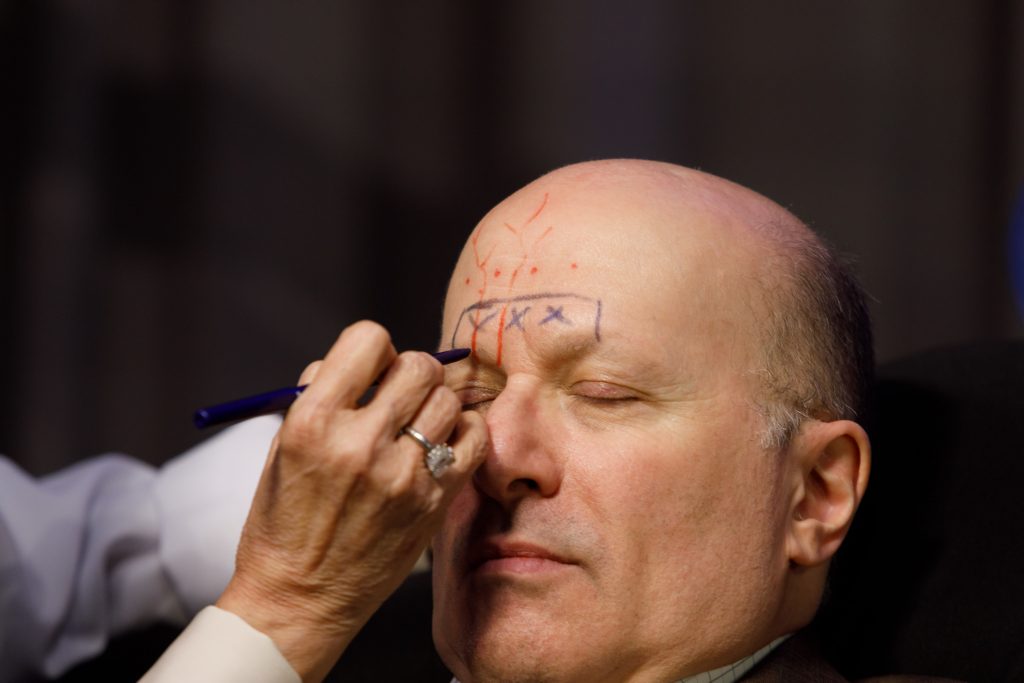
- Injections within 1.5cm of the superior orbital rim (glabellar and medial eyebrows) should be performed superficially as the vessels lie deep in this area. This minimizes the risk of intravascular injections in this area that have the potential to lead to permanent blindness.
- Superior to the 1.5 cm zone above the superior orbital rim, injections may be kept deeper to avoid vasculature and reduce pain (frontal lines).
Important Landmarks for the Middle and Lower 1/3rd of the Face

- The superficial temporal artery exits anteriorly to the tragus and runs superiorly towards the temple. It becomes more superficial as it traverses over the zygoma and then branches to anastomose with the supraorbital and supratrochlear vessels.
- The area along the temple, where the superficial temporal artery is most vulnerable due to its superficial depth, is considered a danger zone. Keep injections deep in this area while avoiding excess pressure as the bone here is very thin.
- The transverse artery runs across the medial cheek but is an end-vessel with no anastomoses. However, it does supply small branches to the infraorbital area.
- The infraorbital foramen lies about 8-10 mm below the inferior orbital rim along the medial limbus. The infraorbital NVB emerges through this aperture. However, the depth of vessels in this area is variable and for safety, a cannula is preferred when injecting in this region.
- The facial artery lies medial to the masseter and runs deep in a tortuous fashion about 1.5 cm lateral to the oral commissure. It supplies the superior and inferior labial arteries which lie deep in the skin.
- The facial artery gives rise to the angular arteries which run along the nasolabial folds most commonly. However, about 30% of individuals may have angular arteries that lie as lateral as near the infraorbital foramen. For safety, a cannula is preferred when injecting this region.
Key Concepts for Middle and Lower 1/3rd Facial Injections
- Injections over the temple should be kept deep to avoid intravascular injections in the superficial temporal artery. However, avoid applying significant pressure in this area as the bone is very thin and may rupture with excess pressure.
- Injections along the lateral zygomatic arch may be kept deep along the bone and this a low risk area.
- When injecting near the infraorbital foramen as well as the nasolabial groove, a cannula can help decrease the risk of intravascular injections. This is due to the vessels in these areas often having variable depth and location.
Adhering to these simple concepts and establishing a sound foundation in anatomy can help dermatologists become more confident in using injectables safely and effectively to achieve the best cosmetic results for their patients.
This information was presented by Dr. Susan Weinkle at the 16th Annual ODAC Dermatology, Aesthetics and Surgical Conference held January 18th-21st, 2019 in Orlando, FL. The above highlights from her lecture and live demonstrations were written and compiled by Dr Nikhil Shyam. Dr. Shyam was one of the 5 residents selected to participate in the Sun Resident Career Mentorship Program (a program supported by an educational grant from Sun Pharmaceutical Industries, Inc.). Dr. Nikhil Shyam is a PGY-4 resident at Johns Hopkins Dermatology Residency Program.
Did you enjoy this post? Find more on Aesthetic Derm topics here.