
Introduction
We present a case of one of the largest cutaneous horns recorded in the known literature as an opportunity to explore diagnostic considerations and treatment options. Cutaneous horns are common exophytic neoplasms composed of dense keratin that are always secondary to primary lesions, which can be benign or malignant. Due to the variance of the primary lesion, diagnostic biopsies are necessary to rule out a malignant origin. Several case reports of giant cutaneous horns may suggest that a larger size indicates a verrucous origin, although a biopsy is necessary as this association has only been noted in very few cases. If the primary lesion is found to be malignant and extending to the biopsy margins, further treatment is required, whereas a benign origin usually requires no further treatment.
Case Summary
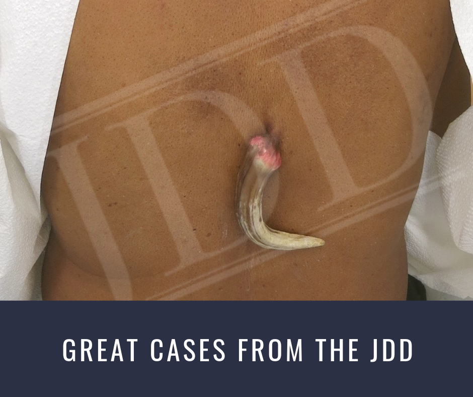
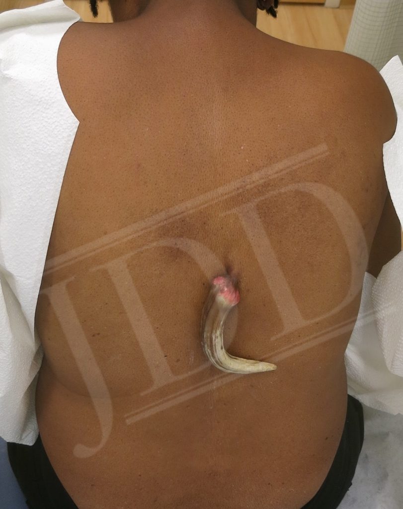
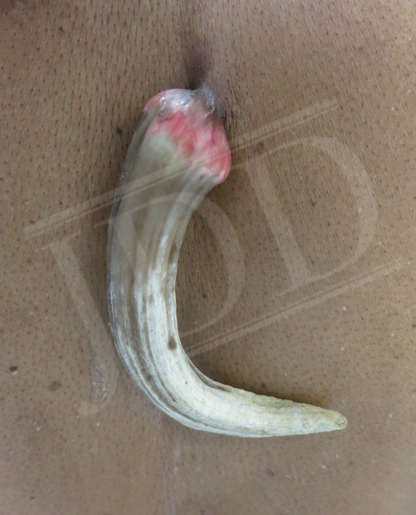
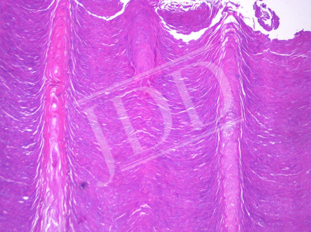
A 65-year-old African American female presented to an outpatient dermatology clinic with an occasionally pruritic and painful “abnormal skin growth” on her back for 22 years. On the mid back was a 22-centimeter tan-brown, horn shaped growth, with a well vascularized base, attached to a pedunculated flesh colored plaque. A shave biopsy was performed below the base of the lesion. Pathology revealed an acanthotic endophytic cellular pattern with protruding columns of parakeratosis, consistent with a cutaneous horn arising from a verruca. Cutaneous horns are compact growths of keratin usually arising from either a verruca, an actinic keratosis, or more concerning, squamous or basal cell carcinoma. In fact, 23% of cutaneous horns develop from malignant cutaneous neoplasms underscoring the importance of histologic analysis and appropriate follow up. Fortunately, the margins on the biopsy specimen were negative/clear and the patient has yet to return with recurrence.
Discussion
A literature search of various synonyms of the words “giant cutaneous horn” suggests that this reported case of a 22-centimeter horn is possibly the largest to ever be described. In looking at similar cases, it appears that cutaneous horns of such a considerable size tend to be secondary to verruca rather than the more dangerous malignant neoplasms.1,2 Although no genetic analysis was performed on this biopsy, one previous case was found to have extensive horn like growths induced by human papillomavirus type two.3 It can be hypothesized that such large horns, and such extensive growth are result of verrucae that may have a genetic predilection in their viral DNA to tremendous angiogenic and proliferative abilities. Cutaneous horns secondary to malignant neoplasms tend to grow in a much more disordered pattern, as would be expected with malignant cells, compared to horns secondary to verrucae.
Cutaneous horns, or “cornu cutaneum” in Latin, are described first in the 16th century, those with horns at the time were used for show. Horns are common among various animals; the main distinction of animal horns and cutaneous horns are animal horns tend to grow off bones giving them strength and cutaneous horns have no bony structure. Similarly, animal horns tend to be common among a species and the same cannot be said for cutaneous horns in humans.4 All human cutaneous horns are pathologic in nature and require a diagnostic biopsy. Again, the differential diagnosis consists of a cutaneous horn secondary to a verruca, squamous or basal cell carcinoma, or actinic keratosis. Further treatment may be required if the origin is malignant and extending to the margins of the biopsy or if the initial biopsy does not resolve an underlying verruca or actinic keratosis. Previous literature may indicate that the larger and more structured a cutaneous horn appears may point to a verrucous, rather than malignant origin but more inquiry needs to be conducted to affirm such an association.
References
- Gould JW, Brodell RT. Giant cutaneous horn associated with verruca vulgaris. Cutis. 1999;64(2):111-112.
- Sanjeeva KK, Ali P, Pinto M, Rao S, Rai AS. Giant cutaneous horn overlying a verruca at an uncommon site: medical marvel vs superstitious dilemma. J Clin Diagn Res. 2015;9(4):PD13-14.
- Chen W, Wei W, Yan-Jun L, et al. Multiple huge cutaneous horns overlying verrucae vulgaris induced by human papillomavirus type 2: a case report. Wiley Online Library. Br J Dermatol. 2018;156(4).
- Bondeson J. Everard Home, John Hunter, and cutaneous horns: a historical review. Am J Dermatopathol. 2001;23(4):362-369.
Source:

Nussbaum, D., BSc, Schwartz, J., MD, & Friedman, A., MD FAAD. (2019). A Giant Cutaneous Horn: One of the Largest Recorded. Journal of Drugs in Dermatology, 18(7), 696-697. Retrieved July 16, 2019, from https://jddonline.com/articles/dermatology/S1545961619P0697X/1/.
Content and images used with permission from the Journal of Drugs in Dermatology.
Adapted from original article for length and style.
Did you enjoy this article? Find more on Derm Topics here.