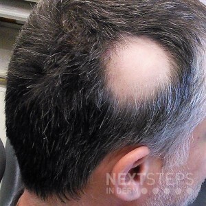
A 36-year-old male presents to the dermatology clinic with the findings depicted in this image. He states that this is the only involved area, and he denies any other associated symptoms. Two 4-mm punch biopsies are performed, one from the affected area and one from the unaffected region. Which of the following histopathologic findings are most likely associated with in this condition?
A. Distorted follicular anatomy, perifollicular hemorrhage, and pigment casts
B. Spongiotic or psoriasiform epidermis, atrophic sebaceous glands, markedly elevated telogen count, and increased miniaturization
C. Peribulbar inflammation with plasma cells, increased miniaturization, and increased telogen count
D. Peribulbar lymphocytic inflammation, dilated infundibula, and increased telogen count
E. Periadnexal lymphocytes, vacuolar interface, and increased mucin
To find out the correct answer and read the explanation, click here.
Brought to you by our brand partner Derm In-Review. A product of SanovaWorks.
![]()
