
During ODAC 2025, Dr. Pooja Sodha — Associate Professor of Dermatology and Director of Laser and Cosmetic Dermatology at GWU School of Medicine and Health Sciences — presented a clinically focused review of the differential diagnosis and stepwise treatment strategies for post-inflammatory hyper‑ and hypopigmentation.
Post-Inflammatory Hyperpigmentation
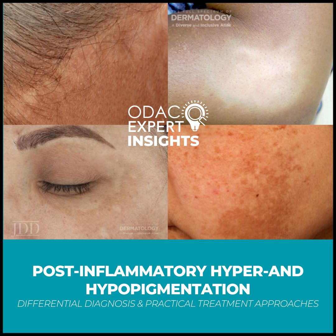
Post-inflammatory hyperpigmentation (PIH) is a pigmentary alteration that occurs due to preceding inflammation, which activates melanocytes and triggers pigment production.
Trauma, wounding, medication-induced reactions, and autoimmune or auto-inflammatory conditions are typical causes. It is important to differentiate PIH from mimickers, like induced pigmentation, drug-induced pigmentation, and melasma.
Identification
The most common ways used to identify PIH is through clinical story and visual examination.
Wood’s lamp (365-nm UV light) can help accentuate epidermal pigmentation from dermal pigmentation. More advanced techniques like parallel/perpendicular polarized photography can be helpful in accentuating different chromophores in the skin, including melanin and oxy/deoxyhemoglobin. Reflectance confocal microscopy can also be used to better delineate the borders of the hyperpigmentation for quantification of the melanin and histological analysis.
Histological Correlation
Epidermal pigmentation indicates accentuation of basal melanocytes and transfer of melanosomes into ascending keratinocytes. Dermal hyperpigmentation reflects injury to basal melanocytes leading to pigment incontinence in the upper dermis. Perivascular leukocytoclastic vasculitis may also be present from the preceding inflammation.
PIH Treatment
Therapies aim to:
-
- Stop the triggering inflammation with antioxidants or anti-inflammatories.
- Target the enzyme pathway that creates melanin.
- Enhance exfoliation and cell turnover with retinoids.
- Block melanosome transfer to keratinocytes.
- Block UV-induced plasmin activation and melanogenesis.
Vascular Component
Dr. Sodha notes that the literature describes between upregulation of vascular endothelial growth factor (VEGF) associated with vascular derangement. Upregulation of VEGF tends to exist in areas of elevated melanin. VEGF triggers an increase in arachidonic acid and prostaglandins which in turn, activates melanocytes, and some melanocytes have direct VEGF receptors to activate melanin production. Addressing erythema early in the treatment of PIH is recommended.
Treatment Algorithm and Options
Dr. Sodha proceeded to present multiple cases of PIH, and the treatments she used in each case. Her algorithm in treatment involves identifying the cause and location of inflammation, considering diagnostic measures, and then targeting the pigment through multiple mechanisms.
Hydroquinone: In one case, she used a milder steroid (desonide) and hydroquinone initially. Hydroquinone is a phenolic compound that inhibits tyrosinase activity. Short duration of use is important to avoid adverse effects.
Timolol: Timolol gel is a beta-adrenergic receptor antagonist typically used in treatment of infantile hemangiomas and rosacea, but she notes good results in use in early abrogation of post inflammatory erythema. It helps with modulating collagen remodeling of the skin as well as fibroblast proliferation. Side effects of hypotension and bradycardia has only been seen in pre-term infants, and PIH treatment calls only for 2-3 drops of 0.5% concentration twice a day for weeks to months.
2 MNG (two mercaptoic glycine compounded with niacinamide): 2 MNG is a non-cytotoxic agent that blocks melanosome transfer by binding to melanin precursors thereby preventing inclusion in eumelanin and pheomelanin synthesis. This will block melanin production while preserving the integrity of melanocytes and the epidermal barrier. As it is not cytotoxic, this therapy may be used long term.
595-nm Pulsed dye laser (PDL): She recommends starting this early (2-4 weeks) as possible in the treatment process to target erythema once active inflammation is quelled. Initial therapy is performed at low pulse durations and very low fluences to for a goal endpoint of very mild erythema, no bruising. Settings are gradually increased in pulse duration and energy over multiple treatments.
Fractional low power 1927-nm diode laser: Used to remove the epidermal pigment that may arise as post inflammatory erythema resolves.
Topical antioxidants and vehicle choice: Dr. Sodha treated a patient with PIH from longstanding acne with topical tazarotene lotion (0.045%) and a compounded topical containing silymarin, vitamin C, ferulic acid, and salicylic acid. Literature shows acne-prone skin has increased levels of oxidative stress and decreased levels of antioxidants. This is thought to be due to lipid peroxidation which occurs in the sebum. Adding a topical antioxidant to the skin can assist in decreasing this process. Silymarin is derived from the milk thistle plant and shown to reduce inflammatory and non-inflammatory acne lesions by 40-45% and improve skin dryness, redness, scaling. Tazarotene lotion formulation enhances tolerability and drug delivery.
Laser assisted drug delivery with tranexamic acid: Dr. Sodha described use of topical tranexamic acid and low-energy, low-density 1927-nm fractional resurfacing laser with laser-assisted drug delivery (LADD) of tranexamic acid immediately post-laser to treat PIH due to chronic atopic dermatitis on the lower anterior legs now controlled on dupilumab. Tranexamic acid reduces plasmin-induced activation of keratinocytes and may competitively inhibit tyrosinase. It is also shown to reduce melanin production in laser-treated cells. Her regimen involves use of the topical prior to starting laser therapy followed by application immediately after laser therapy (laser-assisted drug delivery LADD). Her rational for use 1927-nm diode nonablative fractional laser for LADD is that it coagulates to approximately 170 microns. The depth of the epidermis on the shins is about 100 microns, which identifies the laser as the proper laser to penetrate deep enough for treatment. Channels of approximately 180 microns wide that remain open for around 5 hours, allowing for sufficient drug penetration of tranexamic acid (most particles are less than 180 microns). Use of higher energy devices may be suitable for fairer skin types but requires more caution in skin of color.
Cysteamine cream: She notes using this topical to transition off hydroquinone to a more stable, just as effective treatment with less risk of side effects.
1064-nm picosecond full and fractionated beam Pico-toning therapy: Use of this laser in short pulses will shatter pigment with minimal thermal damage. She like to use a combination of full beam and fractionated therapy. Full beam targets dermal melanophages and basal melanocytes, while fractionated beam supports dermal remodeling. Multiple treatments are performed with low fluence, and expectation management are key.
Treatment for Post-Inflammatory Hypopigmentation (PIHpo)
Case example: Laser induced thermal burn with hypopigmentation in patient with tanned skin
PIHpo after laser hair removal
The mechanism of PIHpo after laser hair is likely due to improperly high fluences, incorrect laser wavelength selection, improper cryogen calibration, or treatment on tanned skin, leading to melanocyte destruction.
Clobetasol ointment: This is used initially for 5 days, then immediately switched to tacrolimus ointment.
Bimatoprost: This is a synthetic prostaglandin F2α analog to activate melanocyte migration and increase melanin production.
1550-nm erbium-doped non-ablative fractional resurfacing laser: This may be used with topical laser-assisted drug delivery of bimatoprost solution immediately post-laser. 1550-nm may be used for patients with tanned skin, aiming for depth of penetration below the dermo–epidermal junction and below.
Topical Tacrolimus: Mouse studies showed tacrolimus improved wound contraction and epithelialization and reduced leukocyte infiltration better than clobetasol. Tacrolimus also promotes melanocyte migration, melanin production, and tyrosinase activity at non-mitogenic levels.
Excimer laser: This laser can be used for hypopigmented scars; however, it does require twice to thrice weekly treatments, 9-10 sessions, with 60-70% improvement reported. Pigment decline may occur around 6 months, requiring retreatment. Combining with tacrolimus or bimatoprost has not yet studied but is something to consider in management.
Key Takeaways
-
- A thorough understanding of the inflammatory process and its impact on melanocytes is crucial for managing both PIH and PIHpo.
- Treatment approaches should be tailored to the individual patient, considering their skin type, the cause and depth of pigmentation, and the stage of inflammation.
- Combination therapies, utilizing both topical agents and energy-based devices, are often necessary for optimal outcomes.
- Early intervention is significantly helpful in both PIH and PIHpo.
- Laser-assisted drug delivery can enhance the efficacy of topical treatments.
- Managing patient expectations regarding the duration of treatment and the need for multiple sessions (especially with energy-based devices) is essential.
This information was presented by Dr. Pooja Sodha during the 2025 ODAC Dermatology Conference. The above session highlights were written and compiled by Dr. Kala Hurst.