Learn more about what it’s like to be a dermatologist today and what it takes to transition from residency to being a practicing dermatologist.
Find hands-on clinical pearls to help you care for your patients more effectively, stay up-to-date with the latest research, or stop by often for expert advice from industry leaders and insights on what it takes to be a key opinion leader.
Mastocytosis is a group of disorders characterized by the pathologic accumulation of mast cells in various tissues. One example of mastocytosis is urticaria pigmentosa, which presents with mastocytomas that can cause hives and, when irritated, pruritus. To the authors’ knowledge, they are describing the first case of urticaria pigmentosa without pruritus. The patient had a positive Darier’s sign, stated that they never felt itchy, and denied ever using a topical steroid or antihistamine. Although their patient declined additional testing, patients like this may benefit from a detailed evaluation of their sensory system through both quantitative sensory testing and genetic analysis.
INTRODUCTION
Mastocytosis describes a group of disorders with a pathologic accumulation of mast cells in various tissues. Urticaria pigmentosa is mastocytosis of the skin, which manifests as mastocytomas that can produce hives and pruritus when irritated. To our knowledge, we are reporting the first case of urticaria pigmentosa without any manifestation of pruritus.
CASE REPORT
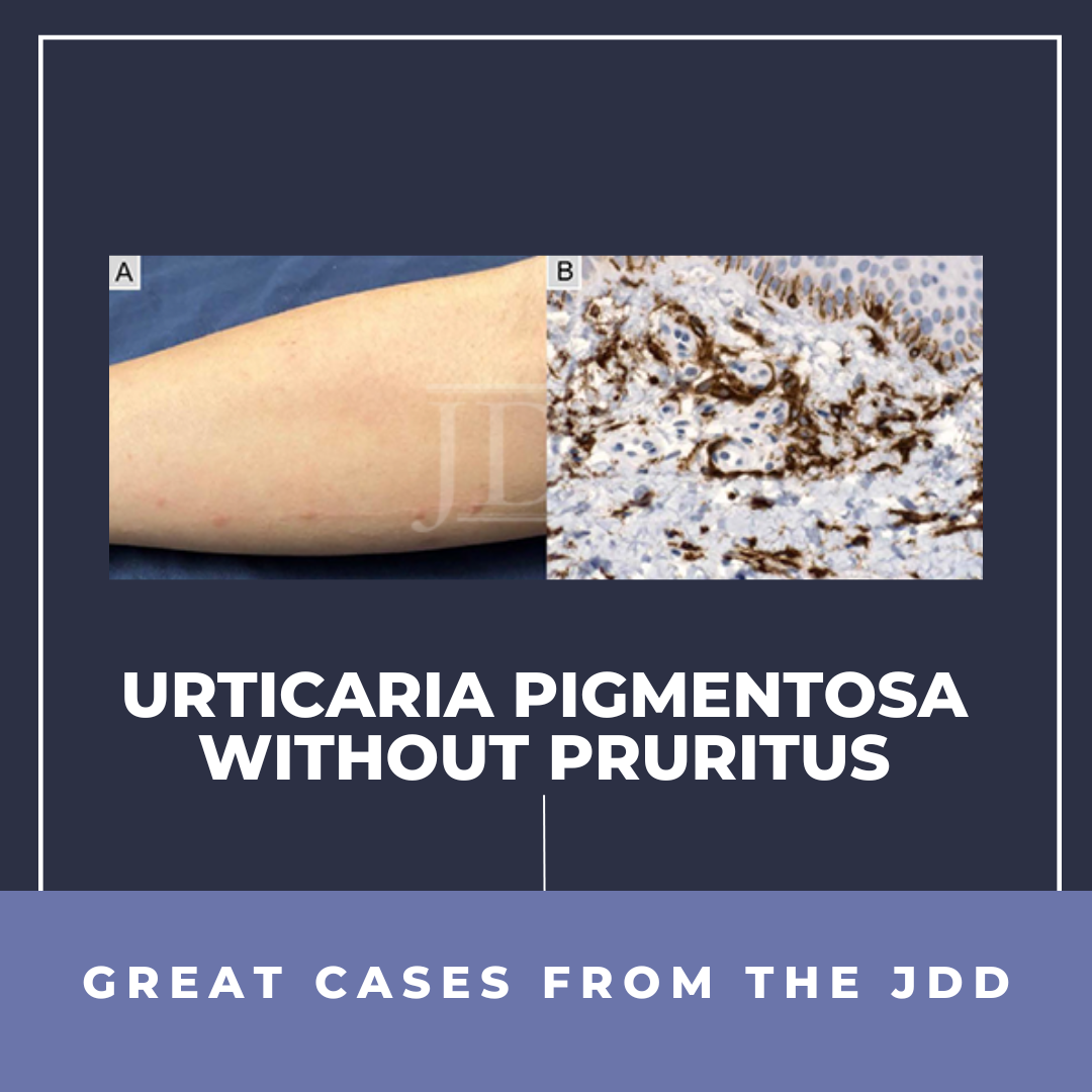
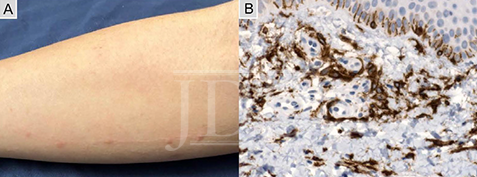
A 27-year-old female presented to the clinic with reddish-brown macules involving all cutaneous surfaces, excluding the face, palms, and soles. Upon stroking the macules on the patient’s forearm, the macules became swollen and erythematous, eliciting a positive Darier’s sign (Figure 1A). A physical examination was negative for hepatomegaly and splenomegaly, but the abdomen was mildly tender. Lymphadenopathy was not detected in the groin, axillae, inguinal, or cervical lymph nodes. Muscle strength was full and symmetric with normal tone and symmetric reflexes. Sensory testing, including vibration sense, was normal. Serum tryptase level was 28 ng/mL (RR, 0-11.4 ng/mL). The patient reported having long-standing episodic bouts of diarrhea and headaches. Moreover, the patient stated that she never feels itching and has never used an antihistamine or topical steroid. A biopsy from a lesion on the forearm demonstrated increased mast cell proliferation. Giemsa staining, immunohistochemical stains for CD117 (Figure 1B), and mast cell tryptase showed 30 mast cells in a high-powered field.
FIGURE 1. Urticaria Pigmentosa. (A) Darier’s sign after stroking areas with mastocytomas on the right forearm. (B) Histopathological specimen from mastocytoma with immunohistochemistry for CD117 showing numerous mast cells (original magnification x400).
DISCUSSION
Mastocytosis is characterized by the pathologic increase of mast cells in tissues, often associated with mutations in the receptor tyrosine kinase KIT (also termed c-KIT or CD117). Mast cells originate from CD34+ progenitor cells in the bone marrow, and they contain a variety of vasoactive mediators that normally function to protect the body via inflammatory responses.1 A mutation in the KIT gene or abnormalities in KIT regulation affect the growth, differentiation, and activation of mast cells.2 In mastocytosis, there is a pathologic activation of the c-kit (CD 117) receptor, leading to unregulated clonal expansion and activation of mast cells. Mastocytosis classically presents with pruritus.
This patient had a positive Darier’s sign, headaches, and loose stools. These signs suggest that mast cells were functioning with active histamine release. The congenital lack of pruritus, with the report of never having used an antihistamine or topical steroids, along with normal sensation on examination suggests that the patient may also have issues with neuronal nociception.
Recently, several human channelopathies involving voltage-gated sodium channels (NaV) have been identified in somatosensory and nociceptive neurons. There are nine NaV channel family members (NaV1.1-NaV1.9) whose functions are determined by the nine distinct pore-forming alpha-subunits. Of these, NaV1.7, NaV1.8, and NaV1.9 have been implicated in itch signaling.3
Pruritogens can activate G-protein-coupled receptors located on nerve endings of primary sensory neurons resulting in a rise in calcium levels. The subsequent membrane depolarization leads to NaV opening and transmission of itch. In patients with congenital insensitivity to pain, the NaV1.7 mutation leads to human pain insensitivity and deficits in itch and temperature discrimination.4 It was found that NaV1.8-/- knock-out models demonstrate impaired histamine and serotonin pruritic scratching.3 There is evidence that a gain-of-function mutation in NaV1.9 alters sensory information from the periphery to the spine, causing debilitating itch and altered pain signaling.5 Thus there appears to be a primary role of NaV1.7 and a contributory role of NaV1.9 in pruritic scratching. Time course studies suggest that NaV1.8 is responsible for prolonged pruritus.3 Mutations leading to such channelopathies, differential signaling expression, and/or altered anatomical expression of NaV1.7, NaV1.8, and NaV1.9 could present with a phenotype of nonpruritus.
In such cases as the authors’, patients should have a detailed evaluation of their sensory system through quantitative sensory testing as well as genetic analysis. Their patient declined to have these.
DISCLOSURES
The authors have no conflicts of interest to declare.
REFERENCES
Metcalfe DD. Mast cells and mastocytosis. Blood. 2008;112(4):946-56.
Bibi S, Langenfeld F, Jeanningros S, et al. Molecular defects in mastocytosis: KIT and beyond KIT. Immunol Allergy Clin North Am. 2014;34(2):239-62.
Kuhn H, Kappes L, Wolf K, et al. Complementary roles of murine NaV1. 7, NaV1. 8 and NaV1. 9 in acute itch signalling. Scientific reports. 2020;10(1): 1-12.
McDermott LA, Weir GA, Themistocleous AC, et al. Defining the functional role of NaV1. 7 in human nociception. Neuron. 2019;101(5):905-19. e8.
Salvatierra J, Diaz-Bustamante M, Meixiong J, et al. A disease mutation reveals a role for Na V 1.9 in acute itch. The Journal of Clinical Investigation. 2018;128(12):5434-47.