
From the SOCU Poster Hall | Pigment-Specific Dermoscopy and AI
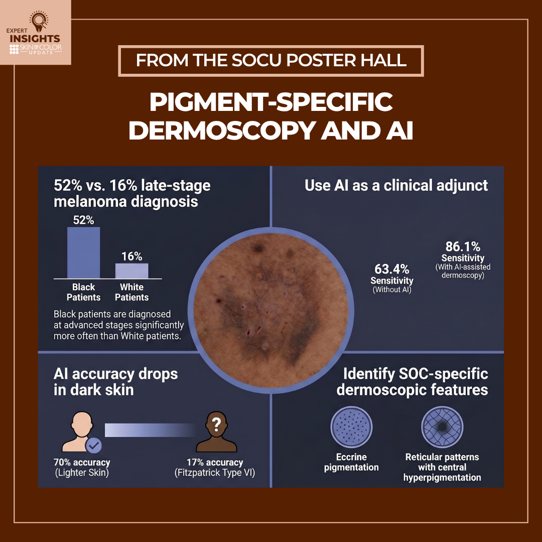 Artificial intelligence (AI) is rapidly changing dermatology, and combining AI with dermoscopy is promising unprecedented diagnostic precision. However, as these technologies advance, a critical question remains: Are they designed to serve all patients equally?
Current diagnostic tools often underperform in patients with skin of color due to biases in training data and a lack of standardized i …
Artificial intelligence (AI) is rapidly changing dermatology, and combining AI with dermoscopy is promising unprecedented diagnostic precision. However, as these technologies advance, a critical question remains: Are they designed to serve all patients equally?
Current diagnostic tools often underperform in patients with skin of color due to biases in training data and a lack of standardized i … Continue reading "From the SOCU Poster Hall | Pigment-Specific Dermoscopy and AI"
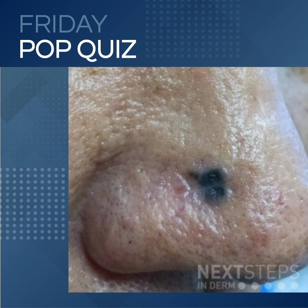 On dermoscopy, which of the following findings most strongly supports the correct diagnosis?
A. Starburst pattern
B. Blue-whitish veil
C. Maple leaf structures
D. Regression structures
E. Irregular streaks
To find out the correct answer and read the explanation, click here.
Brought to you by our brand partne …
On dermoscopy, which of the following findings most strongly supports the correct diagnosis?
A. Starburst pattern
B. Blue-whitish veil
C. Maple leaf structures
D. Regression structures
E. Irregular streaks
To find out the correct answer and read the explanation, click here.
Brought to you by our brand partne … 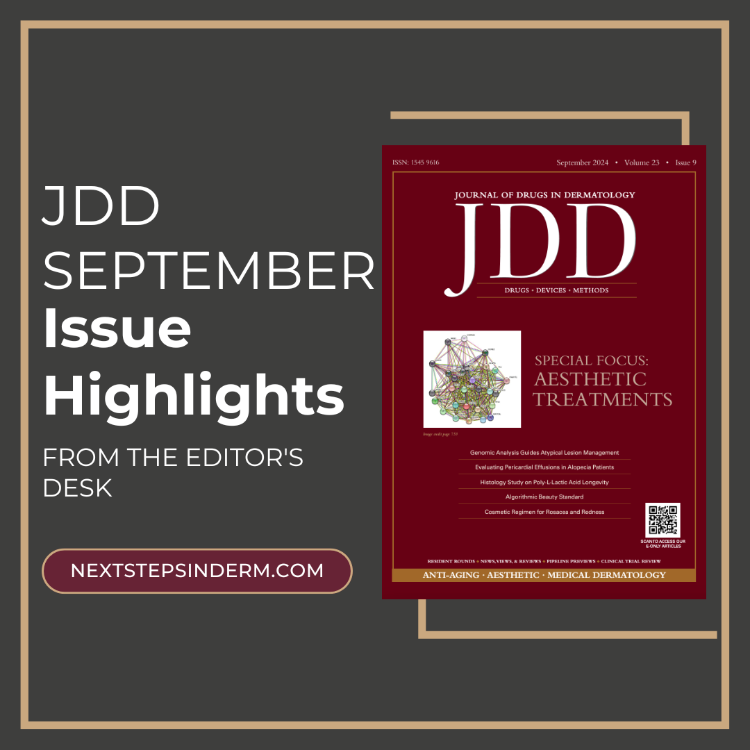 The September edition of the Journal of Drugs in Dermatology (JDD) focuses on aesthetic treatments, exploring the latest advancements and innovative approaches that are reshaping dermatologic care. This month’s Editor's Picks bring together a selection of cutting-edge research, from the use of genomic analysis in melanoma management to AI’s influence on beauty standards.
Explore the latest …
The September edition of the Journal of Drugs in Dermatology (JDD) focuses on aesthetic treatments, exploring the latest advancements and innovative approaches that are reshaping dermatologic care. This month’s Editor's Picks bring together a selection of cutting-edge research, from the use of genomic analysis in melanoma management to AI’s influence on beauty standards.
Explore the latest …  During the 2023 Skin of Color Update in New York City, Dr. Omar Ibrahimi, a renowned laser and cosmetic dermatologist, as well as a Mohs surgeon in private practice in Stamford, Connecticut, imparted valuable insights into the use of lasers for pigmented lesions and tattoos. Dr. Ibrahimi placed significant emphasis on ensuring both safety and efficacy, particularly in individuals with diverse skin …
 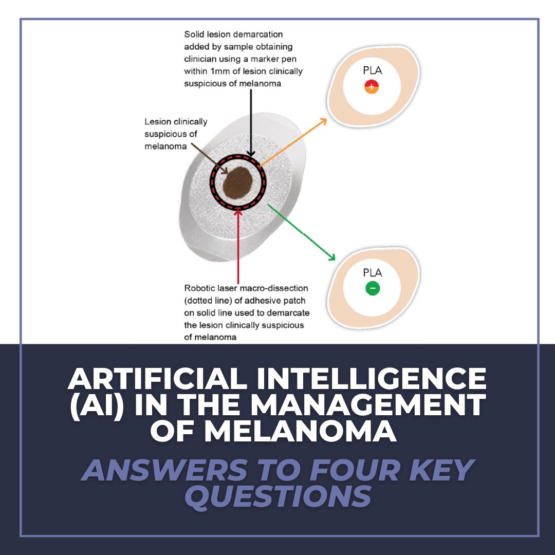 Admittedly, it took me a while to get over the fear of an artificial intelligence (AI) “apocalypse”, which likely developed after my older brother forced me to repeatedly watch “The Terminator” at the tender age of seven. Through an extensive dive into the literature and numerous lectures by Dr. Vishal A. Patel, I’ve since realized the applicability and patient benefit of AI in dermatolo …