
Histologic evaluation of tissue can often be an essential partner to the clinical evaluation in reaching an accurate diagnosis of skin disease. However, clinical pathologic correlation is key for this partnership to be helpful. At ODAC 2022, Dr. Olayemi Sokumbi, dermatologist and dermatopathologist extraordinaire, highlighted this challenge with striking cases demonstrating potential pitfalls and thoughtful pearls that we can all take back to our practice.
“The Double Whammy”
A true pitfall of the clinical pathologic correlation, or CPC, is if both the clinical diagnosis and the pathologic findings both have the same diagnostic mimics. This is what Dr. Sokumi has coined “the double whammy”. To illustrate this lesson, she discussed a striking case of a 28-year-old male with history of positive ANA that presented with indurated erythematous plaques on the extremities, suggesting strongly of connective tissue disease. The biopsy had features of interface, mucin deposition, and lymphocytic inflammation of the fat, which supported a diagnosis of cutaneous lupus. In this case, the clinical impression presented like lupus and so did the histologic features. However, the patient did not respond to classic immunosuppressive treatment, and his skin lesions went on to progress and ulcerate. Repeat biopsy showed a predominant atypical T-cell infiltrate with increased CD8+ expression and TCR gamma delta expression. Now, the diagnosis is cutaneous gamma/delta T-cell lymphoma, which unfortunately is notoriously aggressive and associated with poor outcomes. Dr. Sokumbi reminded us that patients with autoimmune disease are at increased risk of developing lymphoma and further complicating matters. Some lymphomas can mimic autoimmune disease during the initially indolent phase.
“Missing The Unknown”
Another potential for pitfall in the CPC is simple lack of awareness of specific clinical variants of common diagnoses. To demonstrate this, Dr. Sokumbi recounted a case of a 16-year-old female with erythematous, pruritic papules and macules on the elbows and knees. Due to the distribution, dermatitis herpetiformis was suspected. However, the histologic features, which include prominent dermal edema with superficial and deep lymphocytic inflammation, did not support this clinical diagnosis. Further, the patient had no gastrointestinal symptoms or serologies concerning for celiac disease. While this histologic pattern is classic for polymorphic light eruption, the clinical presentation was not recognized as such. However, it all came together once recognizing the newly described spring and summer eruption of the elbows, a localized variant of PMLE.
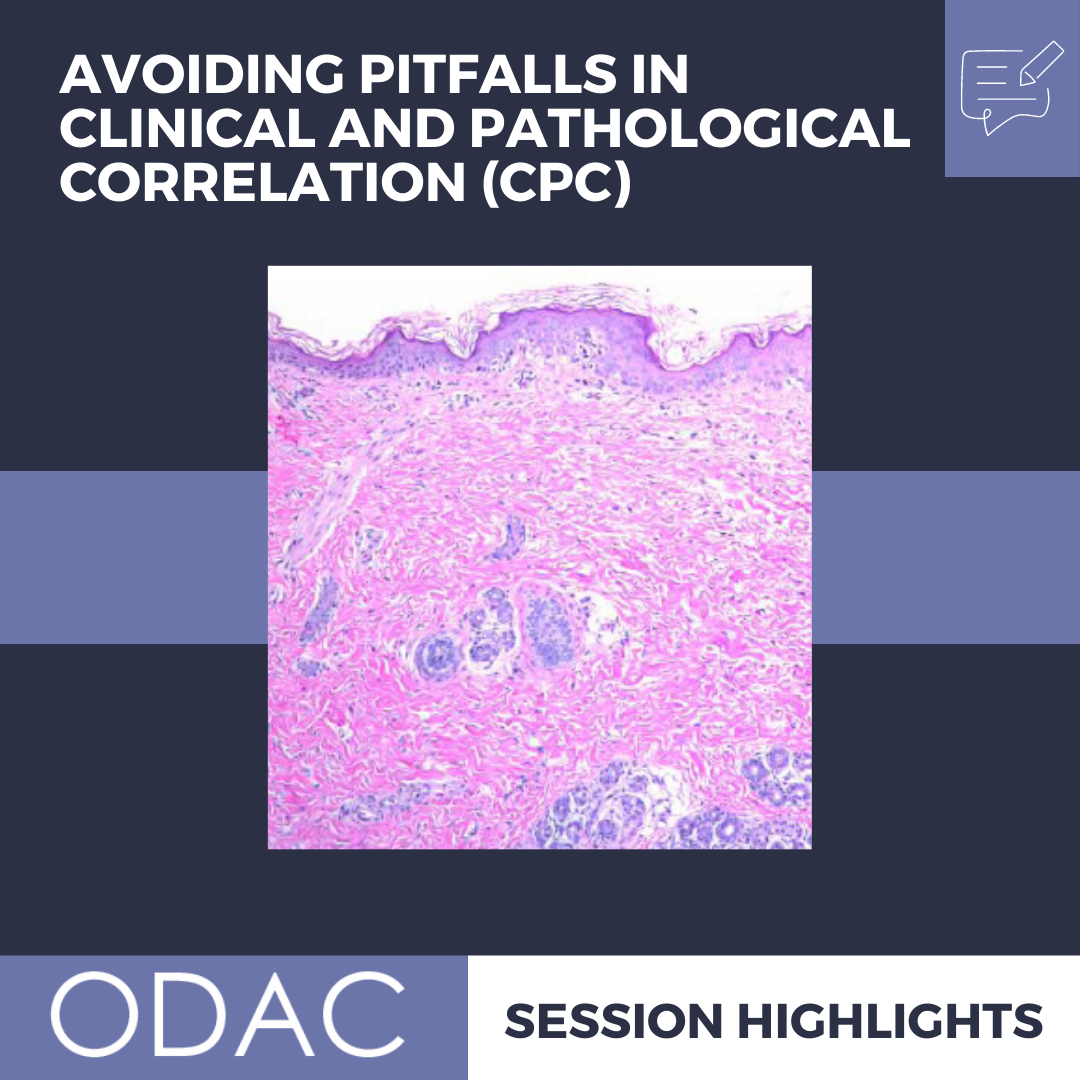
“Staying Superficial”
The dreaded superficially sampled biopsy is a classic pitfall for CPC. What is not shown cannot be evaluated, after all. Dr. Sokumbi utilized a particularly fascinating diagnosis to demonstrate this pitfall, recounting the presentation of a 17-year-old female with asymptomatic, linear hyperpigmented patches on her legs in a peculiar pattern. A shave biopsy only showed evidence of post inflammatory pigmentation, which ultimately was not representation of the diagnosis. A repeat histologic evaluation, of a deeper punch biopsy this time, demonstrated a prominent medium vessel vasculitis in the deep dermis. Strikingly, no neutrophils were present, and the vasculitis was involved only by lymphocytes, which excluded the diagnosis of polyarteritis nodosum. This was consistent with macular lymphocytic arteritis, an uncommon cutaneous vasculitis that lacks systemic involvement.
These striking cases not only brought attention to rare clinical entities in dermatology to our attention, but also demonstrated the importance of CPC to get the right diagnosis for the patients. Dr. Sokumbi reminded us that accurate interpretation of skin biopsy requires an appropriate clinical context. Thus, clinical assessment, thoughtful diagnostic differential and communication with your dermatopathologist are all ultimately key to optimizing the utility of skin biopsies.
This information was presented by Dr. Olayemi Sokumbi at the 2022 ODAC Dermatology, Aesthetic and Surgical Conference held January 14-17, 2022. The above highlights from her lecture were written and compiled by Dr. Edita Newton.
Did you enjoy this article? You can find more on Medical Dermatology here.