
INTRODUCTION
Mohs micrographic surgery (MMS) is increasingly utilized for treatment of skin cancer, however the technique used is markedly different than other surgical modalities.1,2 Explaining MMS to patients is difficult, and anxiety following a skin cancer diagnosis likely leads many to seek out additional resources to supplement their understanding.3,4 It is unclear how accurate and comprehensive these resources are.
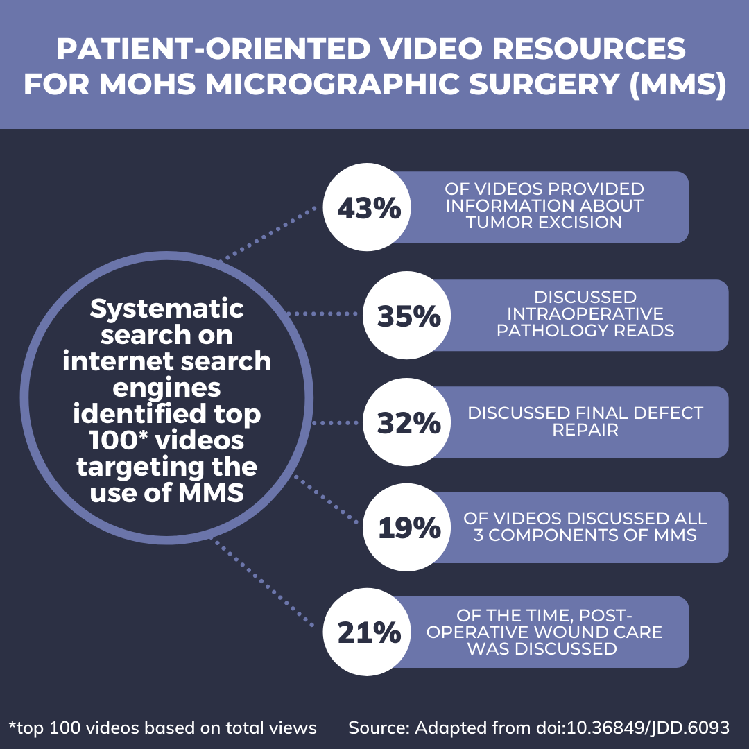
A systematic search was performed on July 18, 2020 and utilized internet search engines to identify videos targeting the use of MMS. Search terms used included: Mohs micrographic surgery, dermatologic surgery, and skin cancer. The top 100 videos based on total views were assessed by 2 reviewers for accuracy and completeness of their descriptions of the MMS procedure, risks and benefits, and post-procedural care. Statistical analysis was performed using Fisher’s exact tests, with P<0.05 considered statistically significant.
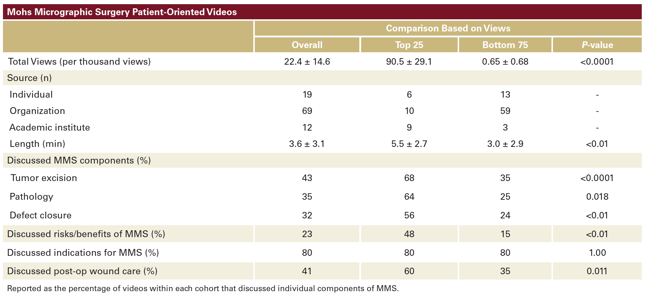
Materials were created by health organizations (69%), individual providers (19%), and academic institutions (12%; Table 1). Average video length was 3.6±3.1 minutes and 78% were created in 2015 or later. Indications for MMS was discussed in 80% of videos, however risks and benefits of MMS were only discussed 23% of the time. Overall, 43% of videos provided information about tumor excision, 35% discussed intraoperative pathology reads, and 32% discussed final defect repair. Only 19% of videos discussed all 3 components of MMS. Post-operative wound care was discussed 21% of the time (Table 1).
The 25 most viewed videos were longer (5.5 ± 2.7 vs 3.0 ± 2.9 minutes; P<0.01), more likely to discuss details of tumor excision (68% vs 35%; P<0.0001), intraoperative pathology (64% vs 25%; P=0.018), and defect closure (56% vs 24%; P<0.01) when compared to the bottom 75. Similarly, they were more likely to discuss risks and benefits of the MMS procedure (48% vs 15%; P<0.01) and post-operative wound care (40% vs 15%; P-0.011).
Ensuring patients understand what to expect during MMS may help reduce anxiety prior to and during their procedure. Most videos neglected the defect repair component entirely, and when it was discussed, it was often too simplified to provide an honest assessment of the likely post-operative wound.
This current analysis highlights the importance of accurately explaining MMS at the time of skin cancer diagnosis, as this is when patients are likely to research the procedure. Further, the creation of comprehensive patient-oriented resources for MMS is needed, as current resources are often insufficient.
DISCLOSURES
REFERENCES
2. Reeder VJ, Gustafson CJ, Mireku K, et al. Trends in Mohs surgery from 1995 to 2010: an analysis of nationally representative data. Dermatol Surg. 2015;41(3):397-403.
3. Callaghan DJ, 3rd. Use of Google trends to examine interest in Mohs micrographic surgery: 2004 to 2016. Dermatol Surg. 2018;44(2):186-192.
4. Tolkachjov SN, Brodland DG, Coldiron BM, et al. Understanding Mohs micrographic surgery: a review and practical guide for the nondermatologist. Mayo Clin Proc. 2017;92(8):1261-1271.
SOURCE
Content and images used with permission from the Journal of Drugs in Dermatology.
Adapted from original article for length and style.