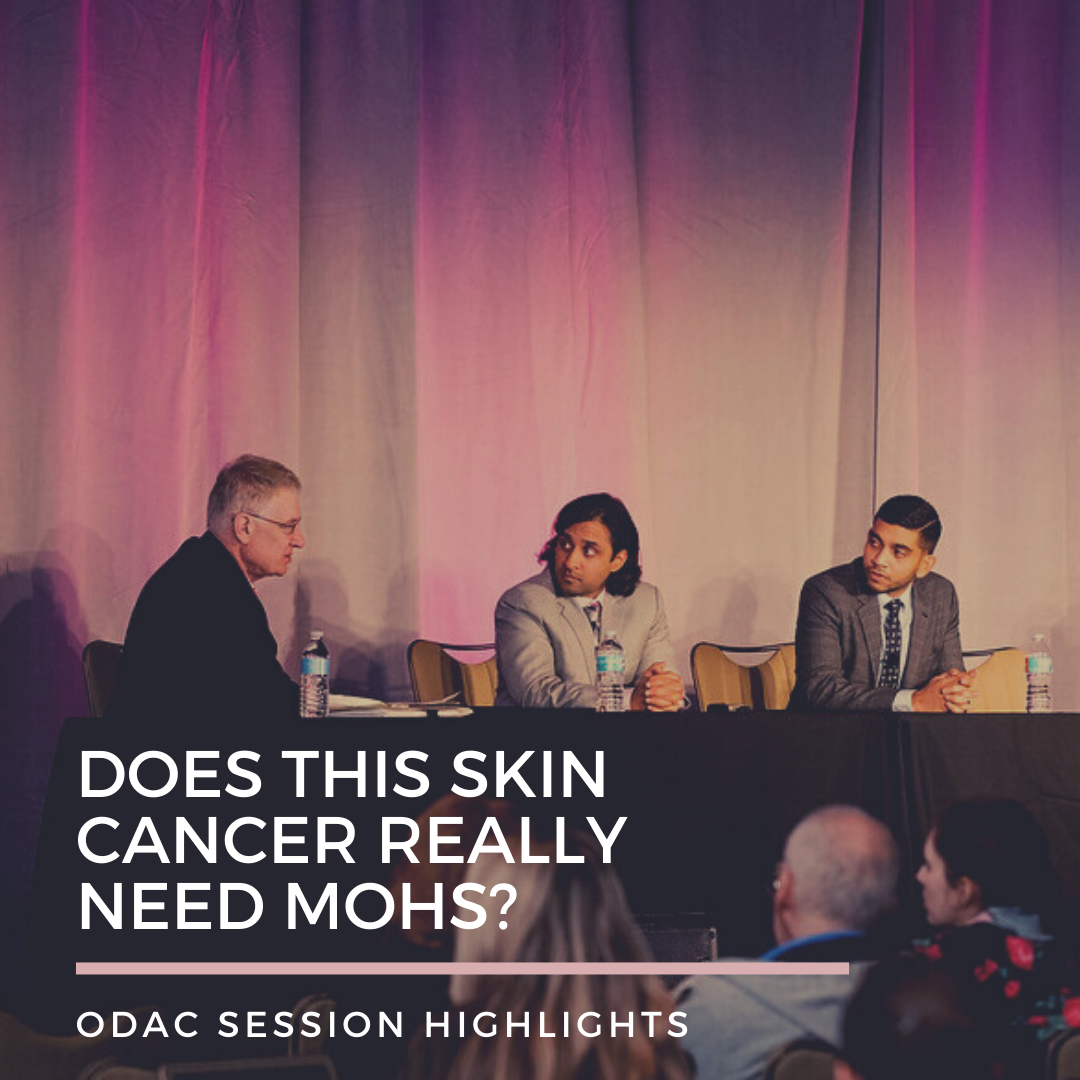
Have you ever come across a patient with a skin cancer that you are not 100% sure should be treated with Mohs surgery or an alternative modality? Or a patient who comes back for a follow-up after skin cancer surgery with an undesirable scar and you wonder if you should have opted for a less invasive option? I certainly have. Some of these nagging questions were answered by a thoughtful, case-based discussion led by Dr. Vishal Patel and Dr. Sailesh Konda and moderated by Dr. William Hanke  regarding the use of Mohs micrographic surgery (MMS) versus other treatment modalities for non-melanoma skin cancers, Merkel cell carcinoma, and lentigo maligna. This panel discussion took place at the 17th Annual ODAC Dermatology, Aesthetic and Surgical Conference in Orlando, Florida.
Case 1
The first case described a 41-year-old Fitzpatrick type II female with two superficial basal cells on her right central cheek (5×7 mm) and right chin (6×8 mm). Mohs Appropriate Use Criteria (AUC) score is 7 for both lesions, justifying MMS for both lesions. Pretty straightforward right?
Think again. Dr. Patel implored the use of topical immunotherapy with imiquimod for these lesions given a study by Williams et al. (1). This randomized clinical trial examined the interventions of either imiquimod 5% cream once daily (superficial basal cell carcinoma, 6 weeks; nodular basal cell carcinoma, 12 weeks) or excisional surgery (4-mm margin) on 3- and 5-year success rates. The 3-year success rate was defined as the clinical absence of initial failure or signs of recurrence at the 3-year dermatology review, and Five-year success was defined as 3-year success plus absence of recurrences identified through medical records. The 5-year success rates for imiquimod were 82.5% compared with 97.7% for surgery (relative risk of imiquimod success = 0.84, 95% confidence interval = 0.77-0.91, P < 0.001). These were comparable to year 3 success rates. Most imiquimod treatment failures occurred in year 1.
Case 2
The second case described a patient we see all too often: a healthy 59-year-old male presents with a squamous cell carcinoma (SCC) in situ on his scalp within a background of actinic keratoses. This patient was referred for MMS.
This case discussed the controversy of upstaging biopsy-diagnosed SCC in situ to invasive SCC during excision or MMS. The commonly used shave biopsy may transect a SCC if performed too superficially, which can make the differentiation between in situ and invasive carcinoma challenging. Several retrospective studies demonstrated that anywhere between to 3.5 % to 16.3% of biopsy-diagnosed SCC in situ were actually invasive SCC following excision or MMS (2-4). Most retrospective studies suggest this rate to be on the lower end, so we should consider other treatment options such as topical therapy (imiquimod and 5-fluorouracil), local destruction, fusiform or disc excision, photodynamic therapy, and lasers (CO2 +/- diode for follicular extension) when treating a SCC in situ in immunocompetent patients.
Case 3
Patient is a 67-year-old male with history of kidney transplant and multiple nonmelanoma skin cancers. He presents with a Merkel cell carcinoma (MCC) of the right preauricular cheek and is referred to surgical oncology for a wide local excision (WLE). Is this the correct treatment option?
Traditionally, WLE has been considered the gold standard for treatment of early-stage MCC. Recent studies demonstrated that MMS appears to be as effective as WLE in treating stage I or II MCC, and may be more effective for MCCs on the head and neck (5, 6). The 2019 National Comprehensive Cancer Network guidelines for MCC recommends treatment in order to obtain histologically negative margins and highlights the use of complete circumferential peripheral and deep margin analysis (CCPDMA) when feasible and to balance surgical margins with surgical morbidity. This suggests that one should consider the use of MMS for MCC treatment. Sentinel lymph node biopsy (SLNB) is always indicated for early-stage MCC and should be done prior to or during the margin assessment surgery when possible to avoid disturbance of lymph node structures.
Case 4
A 72-year-old male with a history of skin cancer presenting with a lentigo maligna on the right cheek was referred for MMS.
Lentigo maligna may be treated surgically with either MMS, staged excision (“Slow Mohs”), or WLE. MMS utilizes frozen sections while staged excision and WLE utilize permanent sections. A recent study suggested that surgical excision of lentigo maligna should include at least 9 mm margins instead of the traditional 5 mm margins (7). Walling et al. found staged excision of lentigo maligna is associated with a significantly lower recurrence rate compared to MMS (8). However, the utilization of MMS for lentigo maligna is gaining momentum, especially with the increasing availability of immunostains, such as MART-1, which can help optimize margin assessment (9). Topical imiquimod may also be used to treat lentigo maligna in select patients when surgery is contraindicated.
References:
-
- Williams HC, Bath-Hextall F, Ozolins M, et al. Surgery versus 5% imiquimod for nodular and superficial basal cell carcinoma: 5-year results of the sins randomized controlled trial. Journal of investigative dermatology. 2017;137(3):614-619. doi:10.1016/j.jid.2016.10.019
-
- Eimpunth S, Goldenberg A, Hamman MS, et al. Squamous cell carcinoma in situ upstaged to invasive squamous cell carcinoma: a 5-year, single institution retrospective review. Dermatologic surgery : official publication for american society for dermatologic surgery [et al]. 2017;43(5):698-703. doi:10.1097/DSS.0000000000001028
- Newsom E, Connolly K, Phillips W, et al. Squamous cell carcinoma in situ with occult invasion: a tertiary care institutional experience. Dermatologic surgery : official publication for american society for dermatologic surgery [et al]. 2019;45(11):1345-1352. doi:10.1097/DSS.0000000000001841
-
- Knackstedt TJ, Brennick JB, Perry AE, Li Z, Quatrano NA, Samie FH. Frequency of squamous cell carcinoma (scc) invasion in transected scc in situreferred for mohs surgery: the dartmouth−hitchcock experience. International journal of dermatology. 2015;54(7):830-833. doi:10.1111/ijd.12867
-
- Singh B, Qureshi MM, Truong MT, Sahni D. Demographics and outcomes of stage i and ii merkel cell carcinoma treated with mohs micrographic surgery compared with wide local excision in the national cancer database. Journal of the american academy of dermatology. 2018;79(1):126-134. doi:10.1016/j.jaad.2018.01.041
-
- Shaikh WR, Sobanko JF, Etzkorn JR, Shin TM, Miller CJ. Utilization patterns and survival outcomes after wide local excision or mohs micrographic surgery for merkel cell carcinoma in the united states, 2004-2009. Journal of the american academy of dermatology. 2018;78(1):175-177. doi:10.1016/j.jaad.2017.09.049
-
- Kunishige JH, Doan L, Brodland DG, Zitelli JA. Comparison of surgical margins for lentigo maligna versus melanoma in situ. Journal of the american academy of dermatology. 2019;81(1):204-212. doi:10.1016/j.jaad.2019.01.051
-
- Walling HW, Scupham RK, Bean AK, Ceilley RI. Staged excision versus mohs micrographic surgery for lentigo maligna and lentigo maligna melanoma. Journal of the american academy of dermatology. 2007;57(4):659-664.
-
- Miller CJ, Giordano CN, Higgins HW. Mohs micrographic surgery for melanoma: as use increases, so does the need for best practices. Jama dermatology. 2019;155(11):1225-1225. doi:10.1001/jamadermatol.2019.2589
Disclosures: Dr. Vishal Patel is a speaker for Regeneron, Sanofi, and Castle Biosciences, and he is the founder of the Skin Cancer Outcomes (SCOUT) consortium
This information was presented by Dr. Vishal Patel and Dr. Sailesh Konda at the 17th ODAC Dermatology, Aesthetic and Surgical Conference held January 17th-20st, 2020 in Orlando, FL. The above highlights from their panel presentation were written and compiled by Dr. Yumeng (Marina) Li, PGY-4 dermatology resident at the University of Miami and one of the 7 residents selected to participate in the Sun Young Dermatology Leader Mentorship Program (a program supported by an educational grant from Sun Pharmaceutical Industries, Inc.).
Did you enjoy this article? Find more on Medical Dermatology here.


