
Who’s afraid of the Big Bad Wolves in pediatric dermatology? Whether in clinic or in the wards, we all have some bad wolves we hope not to encounter, and others that do not fail to surprise dermatologists. Here, I share a few of them as presented by Dr. Yasmine Kirkorian at the 2020 ODAC Dermatology, Aesthetic and Surgical Conference, including Mycoplasma-induced Rash and Mucositis (MIRM), Staphylococcal Scalded Skin Syndrome, PHACES Syndrome, and more!
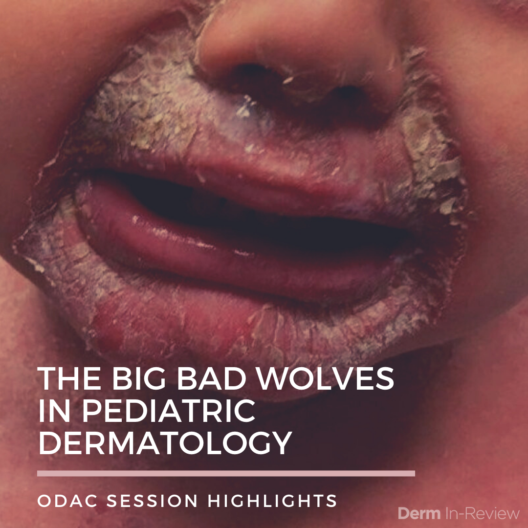
 MIRM or RIME?
Mycoplasma-induced Rash and Mucositis (MIRM) patients are predominantly young males. They almost universally present with a systemic prodrome and have a variable cutaneous eruption:
-
- Mucositis without rash
- Mucositis + scant rash
- Mucositis + extensive rash (SJS-like)¹
MIRM has a good prognosis and should be distinguished from mimickers such as:
-
- Erythema multiforme which is also triggered by an infection, but displays no to minimal mucositis and no skin sloughing, also with good prognosis.
- SJS which is triggered by drugs and displays significant mucositis and skin sloughing.
When there’s no M in MIRM…
Have you encountered a patient with acute onset mucositis, with or without skin rash, no recent drug exposure, had MIRM high on the differential, but mycoplasma PCR was negative?
Here, Dr. Kirkorian presented an 11-year-old with a week of fever, cough, and worsening mucositis. Biopsy showed bullous erythema multiforme. He was treated with IVIG and systemic steroids but showed worsening mucositis and new blisters. On day 11, he was started on cyclosporine A and improved gradually starting the next day. Steroids were tapered and CsA was completed by day 16.
On day 17, new bullae showed, so he was restarted on CsA and methylprednisone. Paraneoplastic pemphigus workup was negative. Again, he improved and was discharged on day 22 on methylprednisone and oral tacrolimus. One year later, he was off all medications and free of symptoms with no recurrences.
Take home points:
-
- Reactive Infectious Mucocutaneous Eruption (RIME) is the broader term when the infection is not mycoplasma.
- It is associated with long hospitalizations, high morbidity, and early treatment may abort the process.
- Consider RIME in any child with abrupt onset mucositis +/- any skin rash
- Treatment: Admission, aggressive pain control, and nutrition. Optimal medications are not yet established, but early CsA can be helpful.
 Staphylococcal Scalded Skin Syndrome
This is caused by toxin-producing strains of Staphylococcus aureus with hematogenous toxin spread. Usually caused by MSSA, which is often clindamycin resistant. Rates are increasing especially in infancy². Importantly, bullous impetigo could be the initial localized source of infection or can be disseminated and overlap with SSSS.
Take home pearls:
-
- In a child with skin pain with perioral and intertriginous erythema, think early SSSS and initiate treatment with antibiotics to reduce desquamation
- Neonates with desquamation: could be SSSS, do not dismiss as physiologic neonatal desquamation.
- Overlap of extensive Bullous impetigo/SSSS can occur
- Swab desquamating lesions, umbilical stump, nares, anus, and circumcision site.
 Who’s Afraid of the Big Bad Bump?
PHACES syndrome:
-
-
-
-
-
- Posterior Fossa Anomalies
- Hemangioma: >5cm and located on the head and/or neck
- Arterial Lesions
- Cardiac abnormalities/coarctation of the aorta
- Eye abnormalities
- Sternal cleft/supraumbilical raphe
-
-
-
-
Pearls for management³:
-
- Echocardiogram, including the great vessels of the neck. Coarctation of the aorta confers the greatest risk of stroke. Obtain echocardiogram before starting propranolol
- MRI/MRA brain/neck/great vessels +/- contrast
- Referral to pediatric neurology and ophthalmology
 Beware the Midline Lesion
The key here is to rule out intracranial communication.
Midline Nasal Lesions in Children:
-
- Nasal gliomas
- Encephalocele
- Dermoid cyst
- Deep infantile hemangioma
- Lymphatic malformation
- Infantile myofibromatosis
- Nodular fasciitis
- Rhabdomyosarcoma
Hence, IMAGE with US then MRI, and if appropriate, subsequent excision to diagnose.
 Who’s Afraid of the Big Bad Fad?
As in all age groups, if it looks like an outside job, it probably is. Below are some of the coolest fads and the not so cool skin presentations:
-
- Slime dermatitis, A Recipe for Disaster
• Borate (Contact lens solution + baking soda, Borax™ detergent)
• Glue
…and for added color and pizazz: food coloring, shaving cream, glitter, or cornstarch - Salt-Ice Challenge, that’s extra cool
• Salt water is used to further lower the freezing point of the ice cube
• Challenge: Keep ice cube on skin as long as tolerated4
• Exam: Geometric/square, areas of erythema, blistering or erosions healing with dyspigmentation and scarring - Deodorant Challenge, smells like trouble
• Challenge: Spray aerosolized deodorant on skin. Watching it frost is
“cool”
• Exam: Cold or frostbite injury with bullae/erosions
- Slime dermatitis, A Recipe for Disaster
Speak with the child privately to gain rapport but expect to not always get a straight answer. Kids do the darndest things…!
References
-
- Canavan TN, Mathes EF, Frieden I, Shinkai K. Mycoplasma pneumoniae-induced rash and mucositis as a syndrome distinct from Stevens-Johnson syndrome and erythema multiforme: a systematic review. Journal of the American Academy of Dermatology. 2015;72(2):239-245.
- Arnold JD, Hoek SN, Kirkorian AY. Epidemiology of staphylococcal scalded skin syndrome in the United States: A cross-sectional study, 2010-2014. Journal of the American Academy of Dermatology. 2018;78(2):404-406.
- Garzon MC, Epstein LG, Heyer GL, et al. PHACE Syndrome: Consensus-Derived Diagnosis and Care Recommendations. The Journal of pediatrics. 2016;178:24-33.e22.
- Yan AC. Current Trends in Social Media-Associated Skin Harm Among Children and Adolescents. Dermatologic clinics. 2019;37(2):169-174.
Image Credits
Clinical image courtesy of Derm In-Review.
This information was presented by Dr. Yasmine Kirkorian at the 17thODAC Dermatology, Aesthetic and Surgical Conference held January 17th-20th, 2020 in Orlando, FL. The above highlights from his lecture were written and compiled by Dr. Saleh Rachidi, PGY-4 dermatology resident at John Hopkins, and one of the 7 residents selected to participate in the Sun Young Dermatology Leader Mentorship Program (a program supported by an educational grant from Sun Pharmaceutical Industries, Inc.).
Did you enjoy this article? Find more on Medical Dermatology here.