
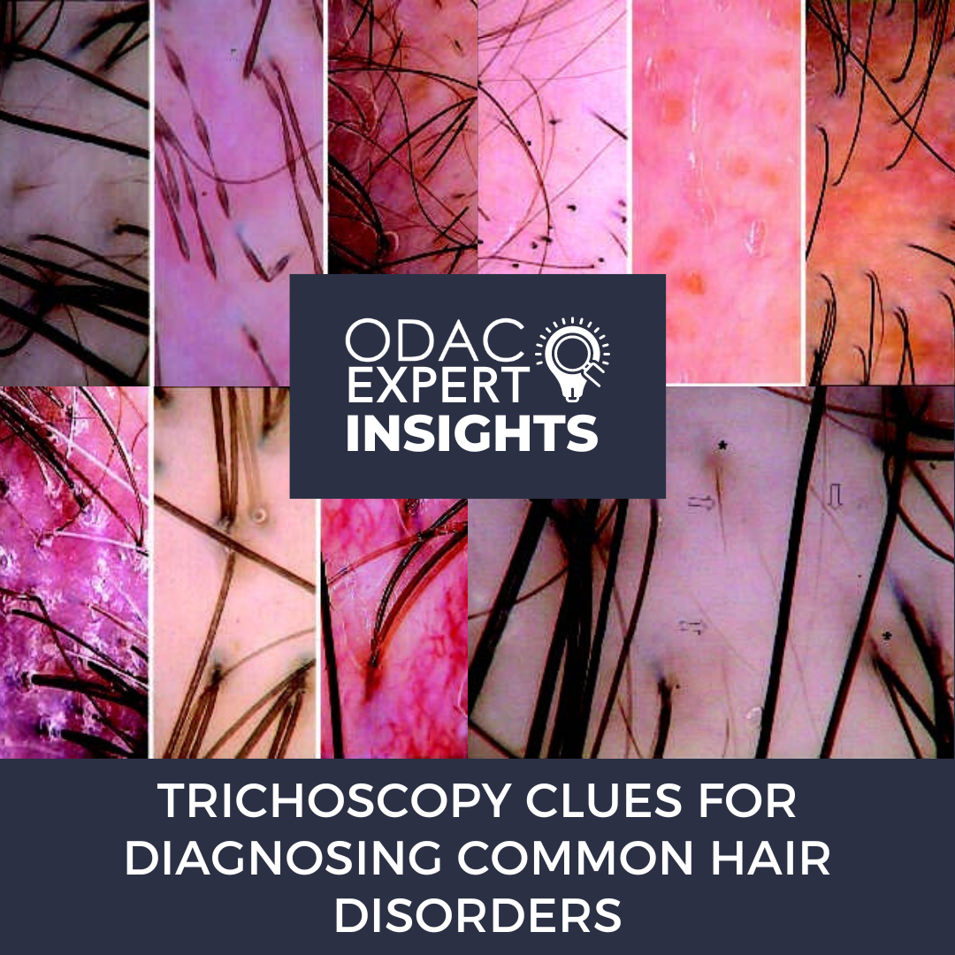
Trichoscopy is a handy dermoscopic tool that can be used at the bedside to diagnose multiple hair diseases. However, these hair diseases may be challenging to diagnose by the untrained eye. Fortunately, we had the opportunity to train these skills at ODAC 2023 with hair expert and dermatologist Dr. Amy McMichael, Professor of Dermatology at Wake Forest University. We will review the essentials of trichoscopy and critical findings in androgenetic alopecia (AGA), alopecia areata (AA), discoid lupus erythematosus (DLE), frontal fibrosing alopecia (FFA), traction alopecia, and central centrifugal cicatricial alopecia (CCCA).
Essentials of Trichoscopy
Trichoscopy can be performed wet or dry on the patient’s scalp. However, when trying to perform dry trichoscopy on the scalp, you should be aware that you may not be able to see scale when the patient’s hair is already wet. This may be because many patients might put oils or moisturizers in their hair before their appointment, which can complicate the diagnostic process. Dry trichoscopy is best for an itchy scalp since with patchy hair loss you can see scale, keratotic plugs, and hair casts. Meanwhile, with wet trichoscopy, you can better visualize vascular patterns of inflammatory conditions using oil. Here are some key locations to look for when performing trichoscopy:
-
- Follicular ostia (where the hair comes out)
- Perifollicular skin (looking for redness, halos, and pigmentary changes)
- Blood vessels (looking for presence or morphology)
- Hair shafts
Androgenetic Alopecia
Case: An elderly woman used color powder to camouflage her hair loss. Now, the patient seeks a better diagnosis to understand the underlying ideology. Trichoscopy revealed hair follicles with terminal hairs and fine hairs. There is also pigmentation around the hair follicles (indicative of sun damage) on the patient’s scalp. There are also sparse fine hairs mixed in. This should help verify the diagnosis of androgenetic alopecia.
Trichoscopy in androgenetic alopecia (i.e., pattern hair loss) shows fine hairs mixed with terminal hairs. There may be an increase in the percentage of single hair follicular units in the frontal area, which may suggest early pattern hair loss. The presence of >6 short, thin hairs in the frontal scalp may be diagnostic. However, also note that short, thin hairs may also be seen in CCCA.
Alopecia Areata
AA reveals exclamation point hairs, dystrophic hairs, broken hairs, and yellow dots in the follicular ostia of empty and hair-bearing follicles on trichoscopy. Looking at the sparse area, you may think of lichen planopilaris. However, the broken and vellus hairs with yellow dots and dystrophic broken hairs should lead you to a diagnosis of alopecia areata.
Trichoscopy Tip: The presence of yellow dots on trichoscopy is characteristic and helpful in diagnosing alopecia areata.
Discoid Lupus Erythematosus
Case: A young woman presents to the dermatology clinic after her hair stylist noticed hair loss at the vertex without thinning. The patient was previously diagnosed with CCCA; however, there is no diffuse thinning on examination. Instead, trichoscopy revealed further clues to the diagnosis; these trichoscopy findings would be unusual with CCCA. Specifically, there is hyperkeratosis with perifollicular scale, areas of hyperpigmentation and hypopigmentation, and follicular dropout.
DLE is a disease seen in all patients, though it is often associated with patients of color. Trichoscopy findings for DLE include a loss of follicular openings, perifollicular scale, and arborizing vessels. There may be sun damage, and keratotic plugs are blocking the follicles. The presence of red dots is common in DLE; meanwhile, the blue-gray dots are usually due to pigment incontinence, which is associated with vacuolar interface changes seen on histopathology.
Trichoscopy Tip: Although pigmentary changes can occur in CCCA, they are often gray rather than the brown changes seen here in the confirmed diagnosis of DLE.
For patients with scale, you may consider other diseases on the differential diagnosis, such as seborrheic dermatitis (since this can be itchy and cause breakage due to scratching). However, the inflammatory changes are helpful in making a diagnosis of DLE. For these patients, shared decision-making should be used to decide on a treatment course, which can include topical corticosteroids, intralesional corticosteroids, and hydroxychloroquine.
Frontal Fibrosing Alopecia
Case: A middle-aged woman presented with hair loss of her frontal scalp that was previously thought to be pattern hair loss. The patient’s occipital scalp appeared normal, and the frontal scalp showed perifollicular scale and erythema. The patient also reports burning and pruritus of her scalp. These findings are consistent with frontal fibrosing alopecia.
Trichoscopy Tip: There may be pattern hair loss associated with frontal fibrosing alopecia, but it may mask the diagnosis without trichoscopy or further evaluation.
Traction Alopecia
Traction alopecia is often the result of trauma on the hair, which may be due to a tight hairstyle. Some patients may not recognize that their hair is tight because they will report that it does not feel tight. Patients may also not come to their appointment with the hairstyle they usually wear, so it is essential to ask them about their usual hairstyle practices. It may be necessary to show these patients dermoscopic evidence of traction alopecia so they know their hair is under tension if it is not feeling tight. Some clinical findings of traction alopecia include the fringe sign—remaining hairs at the marginal periphery of the site of alopecia.
Note that trichoscopy will show the numerous hair casts present in traction alopecia. Hair casts are often a sign of active traction. Perifollicular scale and follicular dropout may be noted as well on trichoscopy of traction alopecia.
Trichoscopy Tip: The flambeau sign, a torch-like shape of hair casts in the direction of pull, is characteristic of traction alopecia.
Central Centrifugal Cicatricial Alopecia
Case: An elderly woman has had central hair loss on the scalp for many years. She presented to the dermatology clinic and wanted to know the cause of her hair loss. The patient previously assumed that it was just age-related hair loss, which may be contributing, but it was not the sole diagnosis. However, a deeper look on trichoscopy revealed some key findings of her disease—CCCA—including:
-
- Peripilar gray/white halos
- Irregular pigmented network
- Perifollicular hyperkeratosis
- Hair shaft variability
In summary, there are many trichoscopic clues to diagnose common hair disorders. Dr. McMichael recommends setting expectations with your patients first, which may include rules that patients should follow to prevent the progression of their diagnosed hair disease. This may include limitations on hair washing, gel or wax use, hairstyle modifications, or other adjustments. With trichoscopy and these five key points, you, too, can be an expert in diagnosing these diseases.
-
- Use your dermatoscope to make a clinical diagnosis.
- Use trichoscopy to focus on the location of your biopsy.
- Check multiple sites for different diagnosis clues (e.g., occipital scalp and frontal scalp).
- Follow inflammation over time using trichoscopy.
- Obtain an attachment for your device to take trichoscopy photos of patient hair (without having to remove your phone cover).
Did you enjoy this article? Find more on hair disorders here.