
Practical Dermoscopy with Sima Jain, MD
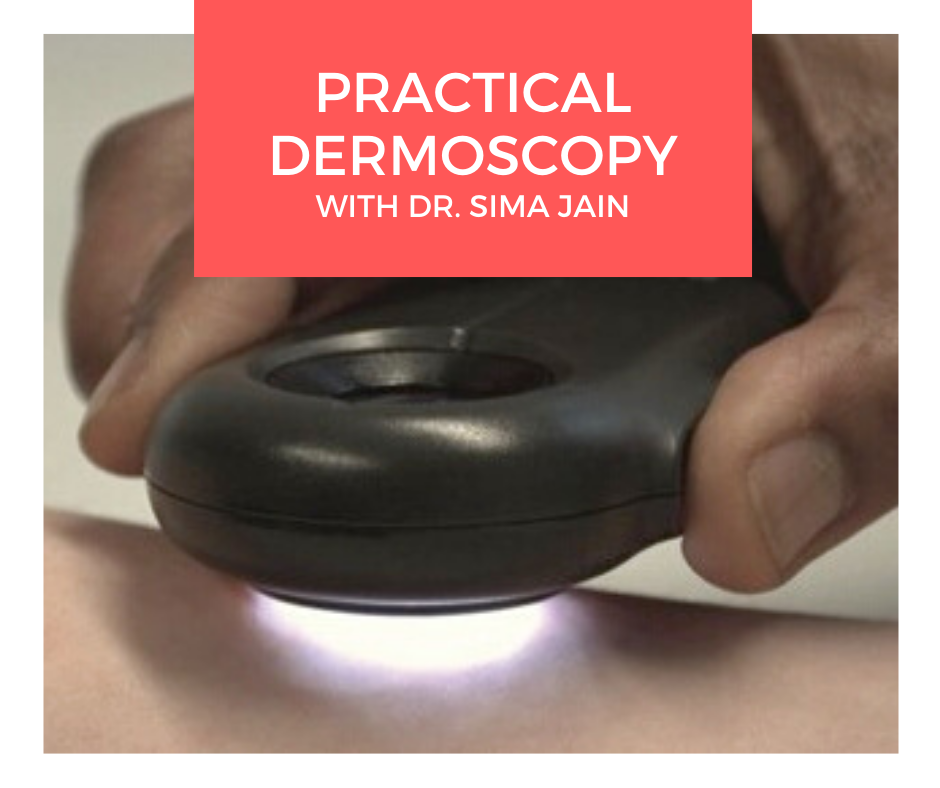 Dermoscopy, also known as epiluminescence microscopy, epiluminoscopy or skin surface microscopy, is an important way to visualize subsurface structures in the epidermis and dermis. In a 2-part series, Dr. Sima Jain reviews the evaluation of pigmented lesions, and the different vessel morphologies and patterns along with a discussion of specific findings in select cutaneous infections.
Read part …
Dermoscopy, also known as epiluminescence microscopy, epiluminoscopy or skin surface microscopy, is an important way to visualize subsurface structures in the epidermis and dermis. In a 2-part series, Dr. Sima Jain reviews the evaluation of pigmented lesions, and the different vessel morphologies and patterns along with a discussion of specific findings in select cutaneous infections.
Read part … 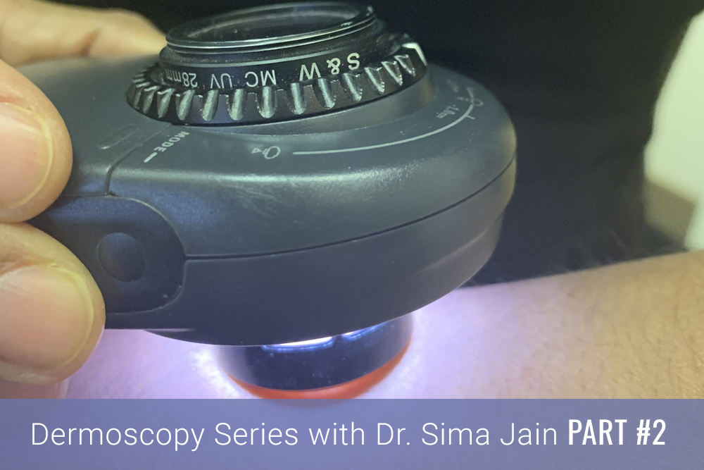 Introduction
Dermoscopy, also known as epiluminescence microscopy, epiluminoscopy or skin surface microscopy, is an important way to visualize subsurface structures in the epidermis and dermis. Part one of this article focused on the evaluation of pigmented lesions, and the second installment below will review the different vessel morphologies and patterns along with discussing specific findings …
Introduction
Dermoscopy, also known as epiluminescence microscopy, epiluminoscopy or skin surface microscopy, is an important way to visualize subsurface structures in the epidermis and dermis. Part one of this article focused on the evaluation of pigmented lesions, and the second installment below will review the different vessel morphologies and patterns along with discussing specific findings … 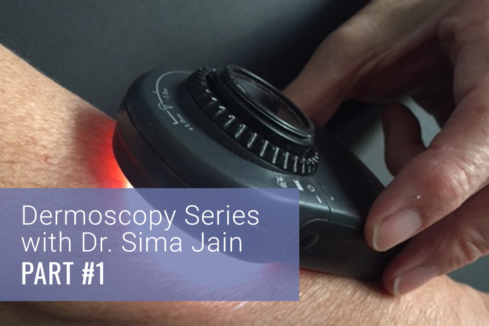 Introduction
Dermoscopy, also known as epiluminescence microscopy, epiluminoscopy or skin surface microscopy, is a noninvasive technique for examination of skin by using a high quality magnifying lens and powerful lighting system to visualize the skin (Figure 1). Although dermoscopy was initially used for the study of mainly pigmented lesions, in the past several years its utility in non-pigmen …
Introduction
Dermoscopy, also known as epiluminescence microscopy, epiluminoscopy or skin surface microscopy, is a noninvasive technique for examination of skin by using a high quality magnifying lens and powerful lighting system to visualize the skin (Figure 1). Although dermoscopy was initially used for the study of mainly pigmented lesions, in the past several years its utility in non-pigmen …