












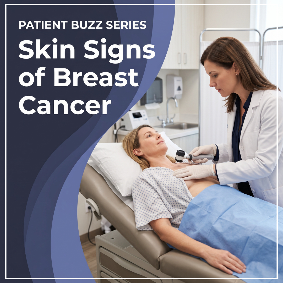
Breast cancer is the most common cancer in women other than skin cancer. Current estimates show one in eight women will develop invasive breast cancer during their lifetime. That means in an average dermatology clinic day, it’s quite likely to encounter a patient who has experienced breast cancer. As noted in an ABC News article, signs of breast cancer can appear on the skin and breast cancer therapies can have skin side effects.
For what to know about breast cancer and the skin, I interviewed Emily R. Nadelmann, MD, FAAD, clinical assistant professor of dermatology at the George Washington University School of Medicine and Health Sciences, and the University of Maryland School of Medicine. Dr. Nadelmann will lecture on complex breast dermatoses at the ODAC Pre-Conference Virtual Symposium. Registration is complimentary.
In your conversations with other dermatology clinicians, do you think breast cancer is often considered when a patient presents with a breast dermatosis?
Yes, I believe it is. It is considered most readily when the presentation matches what we expect: a patient with a known history of breast cancer who develops a new nodule, or a persistent, unilateral eczematous nipple that raises the question of Paget’s disease.
Where consideration becomes less reliable is when the cancer adopts a reaction pattern we associate with something benign. An inflammatory pattern of erythema, edema, and peau d’orange is called cellulitis or mastitis. A sclerotic plaque on the chest wall is read as morphea or radiation fibrosis. A vascular or purpuric pattern in an irradiated breast is attributed to bruising, when it may represent recurrence or radiation-associated angiosarcoma. A firm papule in a surgical scar is assumed to be a keloid. I find it useful to think in these terms: nearly every reaction pattern on the breast has a malignant mimic, and the question is not whether we know breast cancer can involve the skin, but whether we remember to ask which pattern it might be imitating.
What is your clinical experience with patients who presented with a breast dermatosis that turned out to be breast cancer?
The case that comes to mind is a woman in her late sixties who had been treated with lumpectomy and radiation nearly a decade earlier and had been free of disease since. She was referred to me for “keloids” along her surgical scar, a diagnosis that seemed plausible given her history of keloids. Before she reached me, the lesions, several firm, pink, smooth papules within her radiation field, had been injected with triamcinolone on two occasions by another clinician without improvement. What changed my thinking at her first visit was that the lesions had appeared years after her surgery, had continued to enlarge despite those injections, extended beyond the original scar, and felt indurated rather than rubbery. A punch biopsy showed metastatic carcinoma, and the tumor cells were GATA3 positive, consistent with a breast origin. The lesson I took from her is that a scar in a breast cancer survivor deserves the same scrutiny as any new lesion, and that failure of a reasonable treatment is itself a diagnostic finding.
What are the skin signs of breast cancer, including signs that might be overlooked?
The three principal categories are Paget’s disease, inflammatory breast cancer, and cutaneous metastasis.
Paget’s disease presents as a persistent, sharply demarcated, scaly or eroded plaque that begins on the nipple and extends to the areola. It is usually unilateral and may itch, burn, bleed, or accompany nipple retraction. Approximately 85% of cases are associated with an underlying ductal carcinoma in situ or invasive carcinoma.
Inflammatory breast cancer develops rapidly, within 6 months and often within weeks, with erythema, edema, and peau d’orange involving at least one third of the breast. Many patients have no palpable mass, and the erythema may wax and wane. In darker skin, it may appear violaceous, bruise-like, or hyperpigmented rather than red, which matters because inflammatory breast cancer is more common in Black women.
Cutaneous metastases most often present as firm, flesh-colored to pink dermal nodules on the chest wall, frequently in or near a surgical scar. Four less common patterns deserve particular attention because each closely imitates a benign condition:
-
- Carcinoma erysipeloides: a warm, erythematous, sharply marginated plaque that resembles erysipelas or cellulitis. It reflects tumor emboli obstructing dermal lymphatics rather than infection, so fever and leukocytosis are typically absent, and partial improvement with antibiotics can be falsely reassuring.
- Carcinoma telangiectaticum: an erythematous to violaceous patch studded with telangiectasias, hemorrhagic papules, or pseudovesicles that can resemble lymphangioma circumscriptum, vasculitis, or angiokeratomas. Tumor cells may be sparse on biopsy.
- Carcinoma en cuirasse: firm papules and nodules that coalesce into diffuse, woody induration of the chest wall, likened to a breastplate of armor. It is readily mistaken for morphea, radiation fibrosis, or keloids.
- Alopecia neoplastica: smooth, flesh-colored or erythematous patches of scalp hair loss that resemble alopecia areata or a cicatricial alopecia. The distinguishing feature is induration on palpation. Breast cancer is the primary tumor in many reported cases, and the hair loss usually appears months to years after the original diagnosis. Alopecia overlying a presumed scalp cyst should also raise concern.
Other signs that are easily overlooked include new nipple flattening or inversion, subtle unilateral enlargement, isolated nipple crusting, zosteriform papules along a thoracic dermatome, and pigmented metastases that resemble melanoma.
I would add one diagnosis that is not breast cancer at all. In a breast that has been irradiated, new bruise-like or violaceous patches should bring radiation-associated angiosarcoma to mind. It tends to appear 5 to 10 years after treatment, sometimes much later, and it is all too easy to blame on a minor injury or on old radiation change.
What are some other complex breast dermatoses that dermatology clinicians should be aware of?
Nipple adenoma, also known as erosive adenomatosis, is the condition most likely to cause confusion. It presents as an erosive, crusted nipple lesion that is frequently mistaken for Paget’s disease or eczema. Other conditions to keep in mind include hyperkeratosis of the nipple and areola; periductal mastitis and mammary duct fistulas, particularly in young women who smoke; granulomatous mastitis, which can produce peau d’orange and nipple retraction; candidiasis in lactating patients; allergic contact dermatitis to nipple balms, lanolin, or topical antibiotics; and, in irradiated skin, atypical vascular lesions and post-radiation morphea, both of which must be distinguished from angiosarcoma. In patients with breast implants, late swelling, seroma, or ulceration should prompt evaluation for breast implant–associated anaplastic large cell lymphoma or squamous cell carcinoma. Finally, melanoma, basal cell carcinoma, and lymphoma can also arise on the nipple.
When a patient presents with a breast dermatosis, what’s your diagnostic approach?
I begin with the history: laterality, time course, duration, lactation, and any history of breast cancer, radiation, implants, or smoking. I then examine both breasts, both nipples, the axillae, and the supraclavicular nodes, and I palpate the breast itself. For some patients, a dermatology visit is the only clinical breast examination they receive.
When eczema is bilateral and symmetric in a patient with an atopic or contact history, I treat it, but I schedule a defined follow-up within a few weeks. Unilateral or nipple-predominant disease, a sharp border, bleeding, erosion, nipple retraction, or failure to respond to a potent topical corticosteroid warrants a biopsy and diagnostic mammography with or without ultrasound rather than another refill. The biopsy should be a full-thickness skin biopsy of the involved nipple-areolar skin, and the requisition should state that Paget’s disease is a concern. Normal imaging does not exclude Paget’s disease, and breast MRI is the next step when it is confirmed or remains suspected despite a negative mammogram.
For an erythematous, swollen breast with low suspicion of malignancy, a 7- to 10-day course of antibiotics with scheduled reassessment is reasonable. If the breast has not clearly improved, the patient needs prompt imaging and referral to breast surgery. A benign punch biopsy does not exclude inflammatory breast cancer, which remains a clinical diagnosis.
What else should dermatology clinicians know about skin signs of breast cancer?
Delays in diagnosis arise less often from a single missed lesion than from repeated empiric treatment without reassessment. A few habits help: photograph the area at baseline, set a limit on empiric treatment courses, and speak directly with breast imaging when concern is high. In breast cancer survivors, new erythema, induration, nodules, telangiectasias, or bruising within a treated field should be regarded as recurrence or angiosarcoma until proven otherwise, and the oncologic history should always accompany the biopsy, because metastatic cells can be sparse and easily mistaken for inflammation. We should also remember men, in whom Paget’s disease accounts for a larger share of breast cancers and is often treated as a rash first, and we should not wait for redness before considering inflammatory breast cancer in skin of color.
Did you enjoy this Patient Buzz expert commentary? You can find more here.