
Journal Review Series: January-March 2019
 Next Steps in Derm author, Dr. Anna Chacon, searched the journals so that you don’t have to! She reports on important take-aways from different dermatology journals for the months of January, February, and March of 2019.
It is key to keep in mind that “important” is subjective and what is contained in this review is one person’s view of what should be remembered from these months of the …
Next Steps in Derm author, Dr. Anna Chacon, searched the journals so that you don’t have to! She reports on important take-aways from different dermatology journals for the months of January, February, and March of 2019.
It is key to keep in mind that “important” is subjective and what is contained in this review is one person’s view of what should be remembered from these months of the … Continue reading "Journal Review Series: January-March 2019"
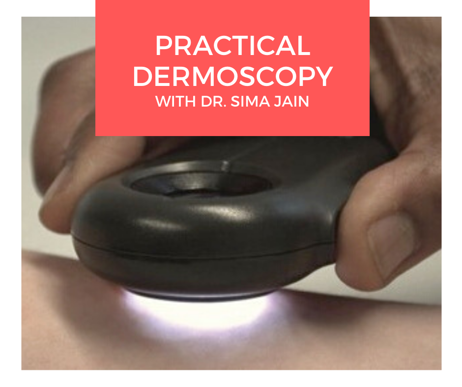 Dermoscopy, also known as epiluminescence microscopy, epiluminoscopy or skin surface microscopy, is an important way to visualize subsurface structures in the epidermis and dermis. In a 2-part series, Dr. Sima Jain reviews the evaluation of pigmented lesions, and the different vessel morphologies and patterns along with a discussion of specific findings in select cutaneous infections.
Read part …
Dermoscopy, also known as epiluminescence microscopy, epiluminoscopy or skin surface microscopy, is an important way to visualize subsurface structures in the epidermis and dermis. In a 2-part series, Dr. Sima Jain reviews the evaluation of pigmented lesions, and the different vessel morphologies and patterns along with a discussion of specific findings in select cutaneous infections.
Read part … 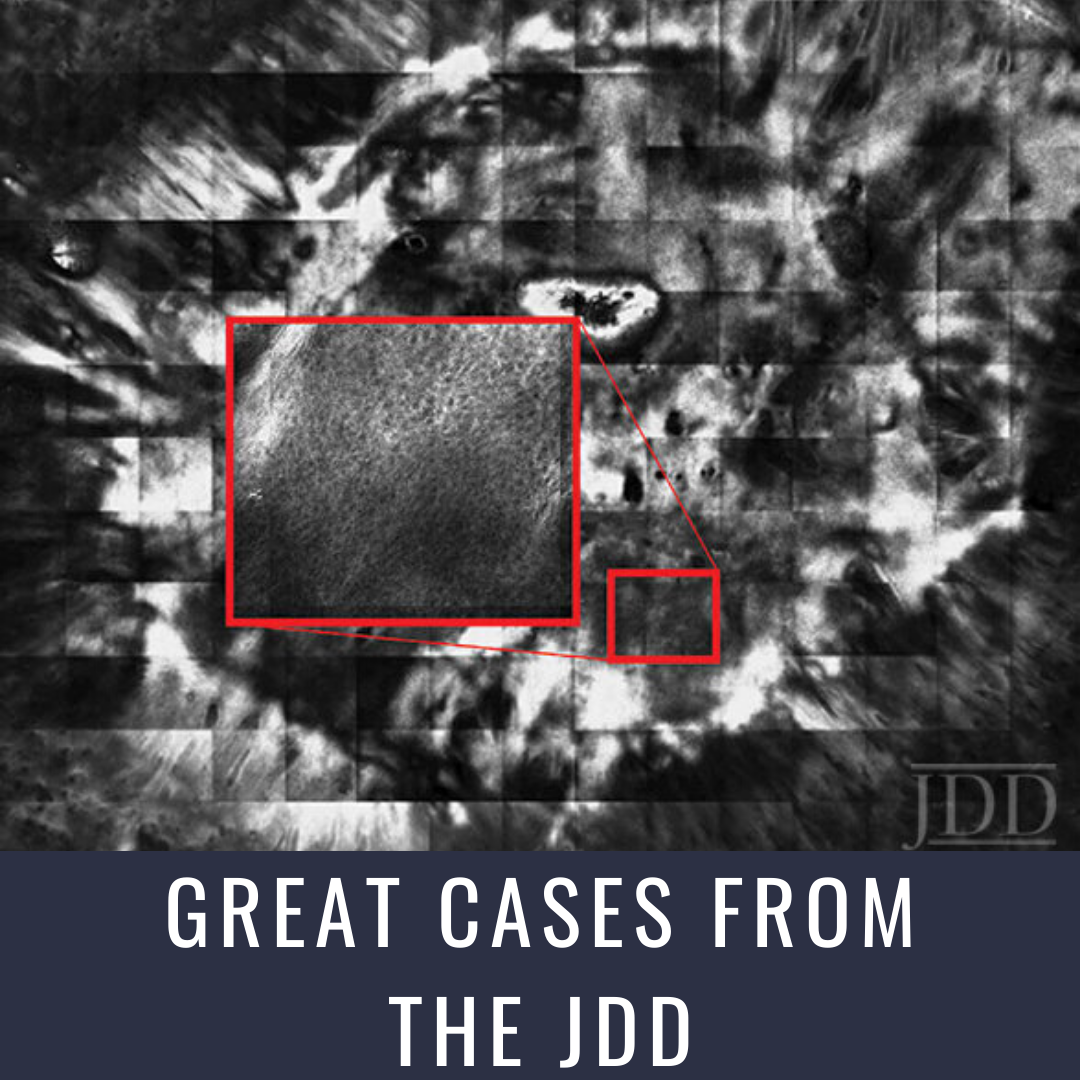 Reflectance Confocal Microscopy (RCM) is a new noninvasive skin imaging modality that is comparable to traditional histopathology. Authors Radhika Srivastava BA, Catherine Reilly BS, Gina Francisco MBS, Hamza Bhatti DO, and Babar K. Rao MD present serial in vivo RCM imaging of an atypical nevus after shave excision over a 1-month period. Findings on RCM images are consistent with the inflammatory, …
 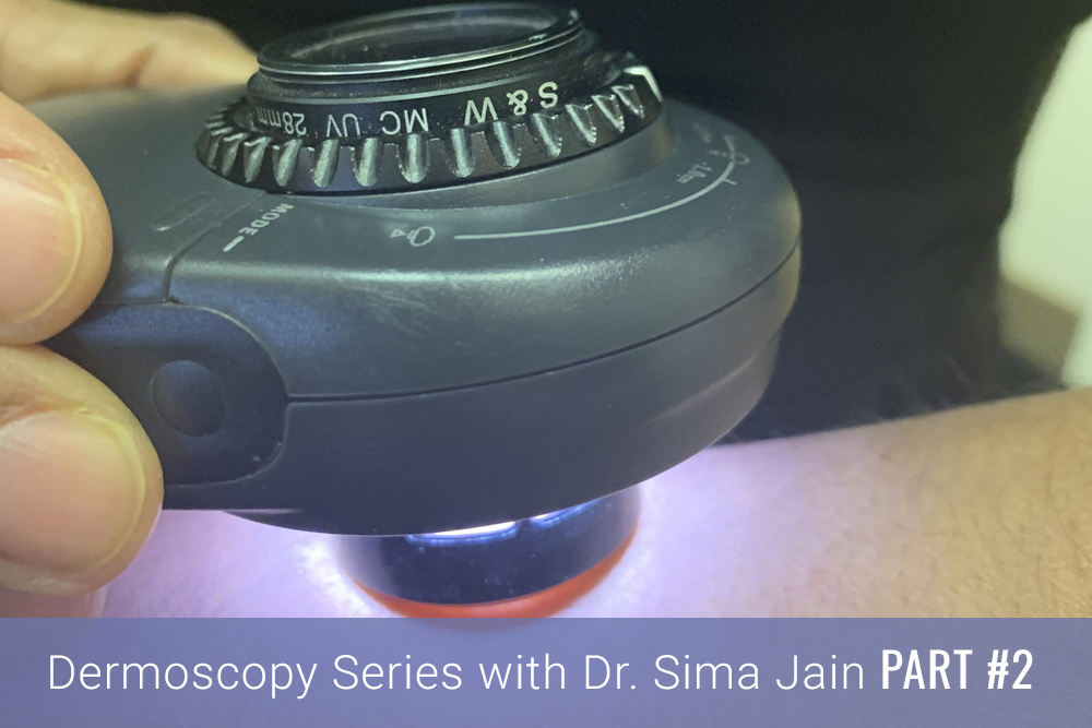 Introduction
Dermoscopy, also known as epiluminescence microscopy, epiluminoscopy or skin surface microscopy, is an important way to visualize subsurface structures in the epidermis and dermis. Part one of this article focused on the evaluation of pigmented lesions, and the second installment below will review the different vessel morphologies and patterns along with discussing specific findings …
Introduction
Dermoscopy, also known as epiluminescence microscopy, epiluminoscopy or skin surface microscopy, is an important way to visualize subsurface structures in the epidermis and dermis. Part one of this article focused on the evaluation of pigmented lesions, and the second installment below will review the different vessel morphologies and patterns along with discussing specific findings … 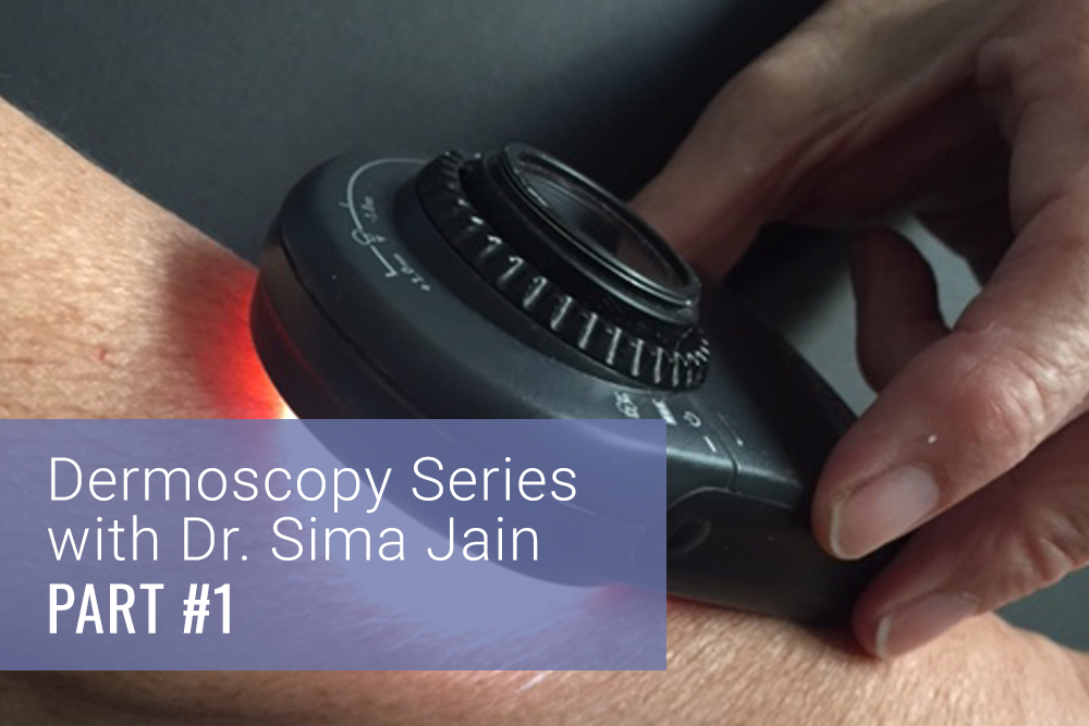 Introduction
Dermoscopy, also known as epiluminescence microscopy, epiluminoscopy or skin surface microscopy, is a noninvasive technique for examination of skin by using a high quality magnifying lens and powerful lighting system to visualize the skin (Figure 1). Although dermoscopy was initially used for the study of mainly pigmented lesions, in the past several years its utility in non-pigmen …
Introduction
Dermoscopy, also known as epiluminescence microscopy, epiluminoscopy or skin surface microscopy, is a noninvasive technique for examination of skin by using a high quality magnifying lens and powerful lighting system to visualize the skin (Figure 1). Although dermoscopy was initially used for the study of mainly pigmented lesions, in the past several years its utility in non-pigmen …