












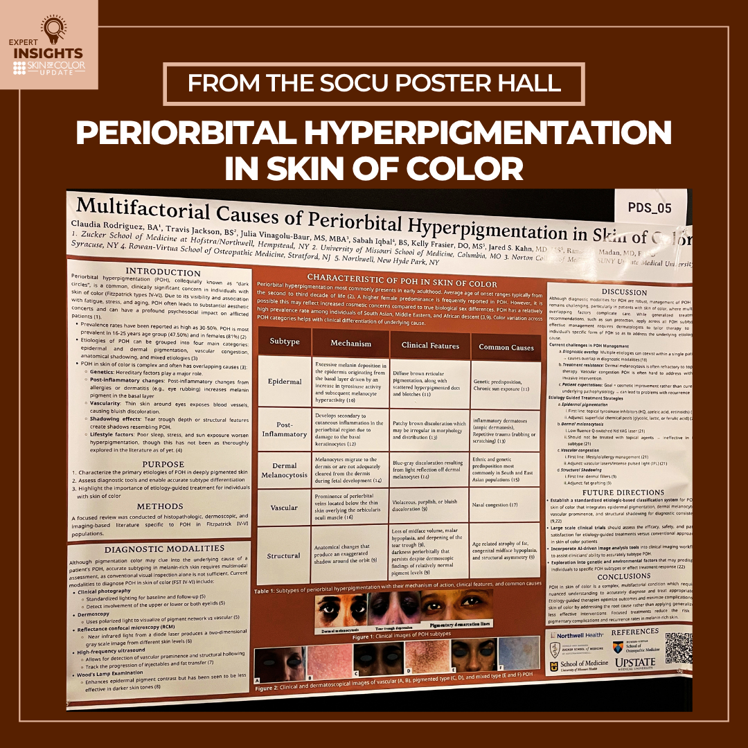
Periorbital hyperpigmentation is common in patients with skin of color, though understanding the condition – and selecting an effective treatment – can be a challenge. Darkening around the eyes can appear as brown, blue-grey, blue, violet, or purple — all signs of the root causes — which include extra deposits of melanin, inflammation, prominent veins, and structural changes. Periorbital hyperpigmentation is not a uniform condition.
The varied etiology of periorbital hyperpigmentation contributes, unfortunately, to poor treatment selection and recurrence. A scientific poster presented at Skin of Color Update looked at the various types of periorbital hyperpigmentation and analyzed the tools that dermatology clinicians can use to make an accurate diagnosis. I interviewed the poster’s presenting author, Claudia Rodriguez, BA, of the Donald and Barbara Zucker School of Medicine at Hofstra/Northwell.
What led you to want to study periorbital hyperpigmentation in deeply pigmented skin?
While periorbital hyperpigmentation (POH) is a common and clinically significant concern in individuals with skin of color, its etiologies and subtypes are poorly delineated, leading to suboptimal results following treatment and increased risk of recurrence. Since POH is estimated to have a prevalence as high as 30-50% in skin of color and can be associated with aesthetic concerns and subsequent psychosocial impacts, it was important for our team to further explore specific etiologies to help inform diagnostics, treatment approaches, and outcomes.
Periorbital hyperpigmentation has a lot of variations in its appearance and causes. What did your study find about the different subtypes of POH?
Our findings discussed that while the etiologies of POH are multifactorial and may be overlapping, they can best be divided into the following subtypes: epidermal, post- inflammatory, dermal melanocytotic, vascular, and structural. Epidermal POH often presents as a diffuse, brown reticular pigmentation and is known to be caused by excessive melanin deposition in the epidermis due to an increase in tyrosinase activity and subsequent melanocyte hyperactivity. Post-inflammatory POH presents as patchy brown discoloration and develops secondary to cutaneous inflammation in the periorbital region due to damage to the basal keratinocytes. Dermal melanocytotic POH presents as blue-grey discoloration and is a result of melanocytes migrating to the dermis during fetal development. Vascular POH presents as violaceous, purplish, or bluish discoloration and is caused by prominent periorbital veins located below the thin skin overlying the orbicularis oculi muscle. Lastly, structural POH presents with a loss of midface volume, malar hypoplasia, and deepening of the tear trough. Here, hyperpigmentation persists despite normal pigment levels and is a result of anatomical changes that produce an exaggerated shadow around the orbit.
Why is it important for dermatology clinicians to categorize a patient’s POH subtype?
Recognition of POH subtype is necessary to drive dermatologists’ therapeutic approaches. While generalized treatment recommendations, such as sun protection, apply across all POH subtypes, effective management requires dermatologists to tailor therapy to the individual’s POH subtype to address the underlying etiologic cause. In this way, dermatologists can set realistic goals and treatment outcomes with their patients, maximizing therapeutic efficacy and durability of response while minimizing both adverse effects and risk of recurrence.
What are some of the diagnostic tools a dermatology clinician can use in determining a patient’s POH subtype, and which tools are most helpful for each subtype?
Although pigmentation can help clue a dermatology clinician into the underlying cause of a patient’s POH, accurate subtyping in melanin-rich skin requires multimodal assessment, as visual inspection may be subjective and is not sufficient as a diagnostic modality. To diagnose POH in skin of color, clinicians may use clinical photography with standardized light, dermoscopy, reflectance confocal microscopy, high frequency ultrasound, and a Wood’s lamp. Clinical photography may be useful for all skin types, while other diagnostic modalities are more subtype specific. For example, dermoscopy is useful in evaluating pigmentation vs vascularity by using polarized light and magnification. RCM, which involves a near infrared light emitted from a diode laser, produces a two-dimensional gray scale so that the location of the pigmentation may be analyzed. Additionally, high frequency ultrasound is useful for detection of vascularity (Doppler) and to assess structural hollowing. It may also be used to track the efficacy of treatment methods such as injectables and fat transfer for structural POH subtypes. Lastly, a Wood’s Lamp examination enhances epidermal pigment, but has been found to be less effective in darker skin tones, making it hard to utilize on Fitzpatrick IV-VI skin types.
How can a dermatology clinician know whether periorbital darkening is caused by pigmentation or shadowing from the structure of the face?
Ultrasound, dermoscopy, and RCM would be useful diagnostic modalities for a clinician differentiating POH caused by pigmentation and that caused by structural hollowing. Through ultrasound, the clinician could utilize Doppler setting to assess vascularity in the region. Additionally, dermoscopy and RCM would allow for the observation of pigment structures or dermal melanocytes that would aid in POH subtyping.
How does knowing a patient’s POH subtype impact treatment selection, and rates of complications and recurrence?
Etiology-guided treatment strategies can help streamline POH treatment in order to achieve quick, long-lasting results that satisfy patients. While patient goals in POH treatment may often be cosmetic, it is important to emphasize curing the underlying pathophysiology to avoid recurrence. By treating the etiology, a clinician can correct the underlying cause of POH while simultaneously avoiding complications and unsuccessful interventions.
Is there anything else a dermatology clinician should know about POH in patients with darker skin tones?
It is important for dermatologists to note that POH etiologies are often overlapping, which can complicate both diagnosis and treatment. When different diagnostic modalities cannot agree on a POH subtype, the patient’s condition becomes more difficult to manage as treatment courses for each individual subtype may be different. Additionally, even when a treatment plan is chosen and adherence is maintained, treatment resistance may occur. For example, dermal melanocytotic POH is often refractory to topical therapy and vascular congestion. POH is often hard to address without invasive interventions. Lastly, treatment of POH often includes the pressure of high patient expectations for cosmetic improvement, regardless of cure for underlying cause. Such a focus on POH’s cosmetic nature can obscure POH as a chronic condition that may in fact relapse. In sum, challenges in management of POH highlight the importance of clear communication between physicians and patients to create a treatment plan that is realistic, sustainable, and ultimately treating the underlying etiology.
Additional authors of the poster include:
Kelly Frasier, DO, MS, Northwell
Travis Jackson, BS, University of Missouri School of Medicine
Julia Vinagolu-Baur, MS, MBA, Norton College of Medicine, SUNY Upstate Medical University
Sabah Iqbal, BS, Rowan-Virtua School of Osteopathic Medicine
Jared S. Kahn, MD, MS, Northwell
Raman K. Madan, MD, FAAD, Northwell
Did you enjoy this article? Find more on Skin of Color Dermatology here.