












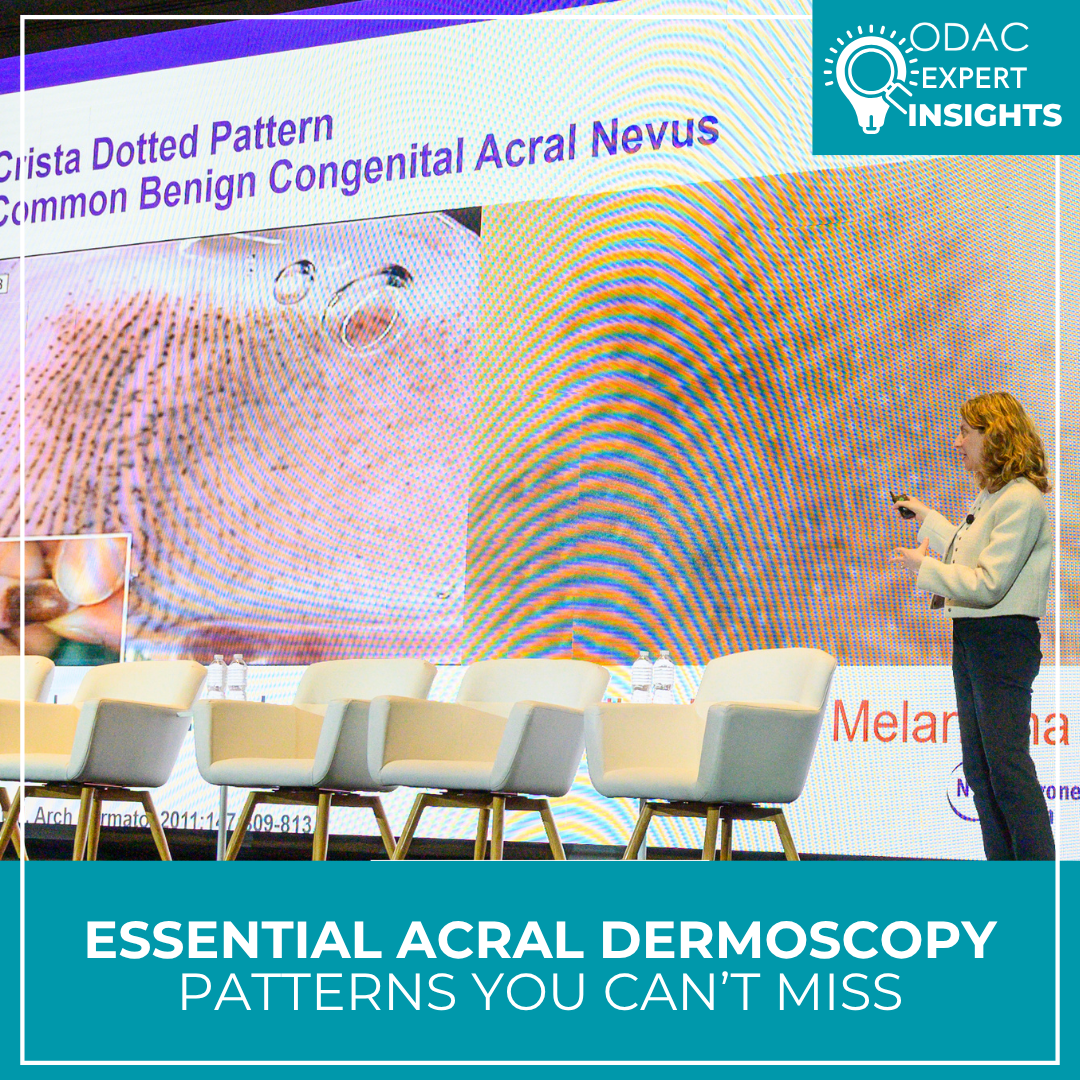
Let’s be honest, acral dermoscopy can be a little scary because no one wants to miss acral lentiginous melanoma. In its early stages, it can be subtle, and outcomes suffer when diagnosis is delayed. In this session, Dr. Stein—professor at NYU School of Medicine where she serves as the associate director of the Pigmented Lesion Section—provides a pattern-recognition reset on volar skin, emphasizing dermoscopic patterns “that we can’t miss and will never miss” after this session, as well as the different “rules” to keep in mind while assessing acral nevi.
#1 The mantra I’m definitely not forgetting
“Furrows are fine. Ridges are risky.”
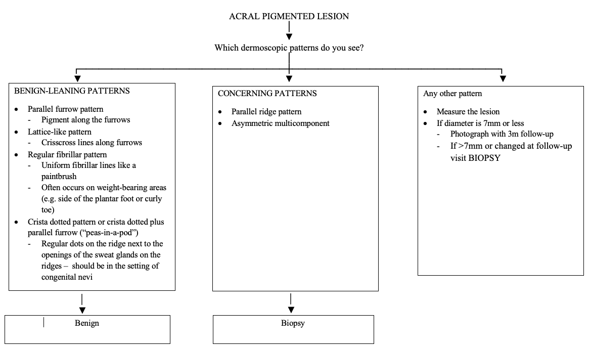
Acral melanoma classically shows pigmentation on the ridges (thick lines, parallel ridge pattern), while pigment in benign acral nevi are more often found in the furrows (thin lines, parallel furrow pattern). There are 3 benign patterns: parallel furrow, lattice-like and fibrillar. Of note, the fibrillar and lattice-like patterns are variants of the parallel furrow. For me, the lattice pattern resembles the grid pattern of a waffle or the crossed strings of a tennis racket. As for the fibrillar pattern, at first glance, it can be confused for a ridge-based pattern since the linear streaks appear to cross the ridges. However, this pattern is caused by shearing forces on weight-bearing skin (e.g side of the plantar foot) causing the pigment to be stretched along the direction of the mechanical force thus forming the fibrillar appearance. However, if the fibrillar lines are not regular, it is more concerning and warrants further evaluation. An easy memory device that Dr. Stein offered for the fibrillar pattern is that it also starts with the letter “f” and are fine (fibrillar = fine).
Clinical Pearl: Furrows = Fine, Ridges = Risky, Fibrillar (regular) pattern = Fine
#2 Patterns you actually need at the bedside
When approaching acral lesions dermoscopically, pattern recognition is key. Below is a simple framework that can guide whether a lesion is appropriate for monitoring or warrants biopsy. However, there are some variants you should keep in mind.
Variants
Acquired
In Fitzpatrick types V-VII skin, you tend to see many of these smudgy, kind of light brown-tan lesions on the bottoms of the feet or hands that seem to have this homogenous, bland ridge-like pattern. These physiologic volar melanotic macules are a common yet neglected pigmentary variant that are also known as acral or ethnic melanosis. If you’ve had a patient of skin of color, you most likely have observed this. However, you shouldn’t just write this variant off because there could be a melanoma in a patient who happens to have this phenomenon, so look for an outlier lesion.
Congenital
In addition to the parallel ridge pattern, the crista dotted pattern is a common pattern seen in congenital nevi. In the crista dotted pattern, histologically, these “dots or globules” are regularly spaced next to the ridges i.e. openings of the eccrine glands (this makes sense since we tend to see melanocytic nevus nests next to adnexal units like sweat glands). It can be seen in combination with the parallel furrow pattern, and if you look close enough, it resembles peas-in-a-pod! This orientation seems “risky” as it involves the ridges but when compared to a melanoma you can see the difference.
Don’t skip nails when you’re “doing acral”
Acral dermoscopy inevitably drifts into nail questions, and the session’s practical point is that dermoscopy can spare patients months of anxiety (or speed the right biopsy):
-
- Subungual hemorrhage often shows well-circumscribed dots/lacunae or homogeneous color, sometimes with distal streaking depending on timing and location.
- Fungal melanonychia tends to be more yellow and dermoscopically has this “reverse triangle” pattern (wider band present distally)
- Ethnic melanonychia presents with longitudinal bands that can be found on multiple nails
-
-
- Normal nail variant
- Can think of it as multiple lentigines present in the matrix
- Should be the same thickness proximally as distally
- However, be on the lookout for a nail with an irregular band with a different width and color and looks different from the rest. This should be biopsied.
-
-
- Nail melanoma risk features include destruction of the nail plate, irregular bands of different colors and thickness, and Hutchinson sign (periungual pigmentation on the skin) and wider at the base than distally.
-
-
- A pseudo Hutchinson sign is where the pigment is found on the nail plate (can be seen through translucent cuticle on dermoscopy) and can be found in benign nevi.
-
Clinical pearl: look for the outlier or ugly duckling when it comes to nails and acral nevi.
This information was presented by Jennifer Stein, MD, PhD at the 2026 ODAC Dermatology Conference. The above highlights from his lecture were written and compiled by Ndidi Enwereji, MD, MPH.