Women’s Health recently wrote an article about four skin conditions that are commonly misdiagnosed. Why do primary care providers sometimes misdiagnose common skin conditions? What conditions are challenging for dermatologists to diagnose? What steps can a dermatologist take to make a differential diagnosis?
For expert advice, I contacted Steve Daveluy, MD, FAAD, associate professor and program director of Wayne State University Department of Dermatology, and president of the Wayne County Medical Society of Southeast Michigan.
Why do primary care providers sometimes misdiagnose common skin conditions?
As Dr. Lee mentions in the article, diagnosis of skin disease is very challenging and it takes years of training for dermatologists to develop the skills to make an accurate diagnosis. Most non-dermatologists have less than a month of dermatology education, so misdiagnosis is not uncommon. General practitioners can’t dedicate the time and effort to extensive training on dermatologic disease. So, it’s important for primary care doctors to recognize when it’s time to send a patient to see a board-certified dermatologist for assessment.
What are the most common misdiagnosed conditions you see in your clinic?
One of the commonly misdiagnosed conditions I see is actinic keratosis. In patients with many actinic keratoses on the face and forearms, physicians may mistake them for a rash. They are red and scaly like a rash.
As Dr. Lee discussed in the article, hidradenitis suppurativa (HS) is often misdiagnosed as folliculitis or simply recurrent abscesses. In the case of HS, it’s actually a pretty easy diagnosis, as long as you know that HS exists. We need to increase awareness of the disease, so physicians recognize it when they see it and refer the patient to a dermatologist.
Stasis dermatitis of the lower legs is a very common misdiagnosis. We get many consults for “bilateral lower extremity cellulitis,” which always turns out to be stasis dermatitis.
Granuloma annulare is often confused for a tinea infection since they are both annular. Nearly every patient referred to me with granuloma annulare has already been treated with a topical antifungal cream due to misdiagnosis as a tinea infection.
What are the most challenging conditions for dermatologists to diagnose and why?
 Mycosis fungoides (MF), the most common form of cutaneous T-cell lymphoma, often presents a lot of diagnostic challenges. It can be difficult to make the diagnosis for a few reasons. Sometimes the rash of MF has all the classic features: bathing trunk distribution, “cigarette paper” atrophy, fine-scale, or poikiloderma. But when it doesn’t have many classic features, it can be very difficult to distinguish from other rashes, especially eczema. To make things more difficult, it doesn’t always show up on biopsy. There are not usually great numbers of atypical T-cells in the skin, so it can be hard to catch them on biopsy and often requires multiple biopsies before you catch it. That’s where the mysterious disease known as parapsoriasis comes into play. When you clinically suspect a diagnosis of mycosis fungoides, but the biopsy is not consistent and shows nonspecific findings, you make a diagnosis of parapsoriasis. You start treatment with topical steroids, and in some cases phototherapy, but you keep an eye on it and consider a repeat biopsy if it’s not responding to therapy. Some cases of parapsoriasis will be diagnosed as mycosis fungoides on repeat biopsy.
Drug-induced rashes can be challenging as well. Many times, the history gives it away: the patient starts a new medication and develops a rash within the first few weeks. But it’s not always so easy. Calcium channel blockers and hydrochlorothiazide can cause an eczematous dermatitis in patients aged 60 and older (mean age 76) that typically presents after taking the medication for at least three months. It eventually improves after discontinuation of the drug, but it can take a year after drug cessation. A drug-induced subacute cutaneous lupus can be caused by proton pump inhibitors, which are available over the counter. Antiepileptics (phenytoin, carbamazepine, valproic acid), gemcitabine, gold, and the clonidine patch can cause a rash that will appear identical to mycosis fungoides on biopsy, even showing atypical T-cells. It’s important to review every patient’s list of medications for possible drug-induced dermatoses.
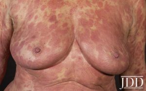
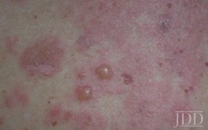
 Bullous pemphigoid can be a challenge when it presents without any bullae. It’s also important to consider bullous pemphigoid in elderly patients presenting with an eczematous dermatitis, urticarial eruption, or just pruritus. It can present without bullae and require biopsy for direct immunofluorescence to make the diagnosis.
Always consider the great mimickers in dermatology, syphilis, and sarcoidosis. They can both present with a wide variety of skin findings.
It’s also difficult to diagnose skin infections that come from other parts of the world. You will have limited to no personal experience with these diseases, but patients may acquire them in other parts of the world and then show up in your clinic.
What are the key steps involved in assembling a differential diagnosis?
When forming a differential diagnosis, you need to consider all the features you see in your patient and compare them to the features seen in different diseases. This includes elements from the history: onset, severity, associated symptoms, exacerbating factors, prior treatments, and responses to treatment; as well as elements from the physical exam: distribution, morphology, and primary and secondary skin findings. Morphology is very important in dermatology and allows you to focus your differential. You can consider broad categories: Do I think this is an infectious process, neoplastic process, or inflammatory process? When diagnosing rashes, certain elements of the morphology allow you to consider more narrow categories such as papulosquamous diseases, dermal inflammatory diseases, vascular diseases, etc. This not only helps you when building a differential from your knowledge, but it also helps when you need to utilize the literature to expand your differential.
In your work with residents, what do you consider to be the most challenging aspect of teaching and training residents on assembling a differential diagnosis?
When teaching residents about building a differential diagnosis, I often talk about the way our brains work. Our brains have two modes: fast thinking and slow thinking. With experience, dermatologists can diagnose common skin problems with fast thinking. You take one look and you know what it is. Our brains love fast thinking because there is such information around us all the time that the brain would become overwhelmed if it didn’t have shortcuts. But when you can’t instantly make the diagnosis, you shift into slow thinking. You spend more time with each feature and put them all together. You may consider each diagnosis you’re considering. For example, a patient’s rash may have the morphology of psoriasis, but not the typical distribution.
One of the challenges in residency and early in your career is building your knowledge of skin diseases. You won’t include a disease in your differential if you’ve never heard of it. As you acquire more knowledge and experience, building a differential becomes easier. It’s a challenge for everyone to remember to consider rare diseases as well since they don’t pop into our heads as readily. That’s also why it’s so important to keep learning throughout our careers.
What other suggestions or guidance do you have for dermatologists in helping them make accurate definitive diagnoses?
When something doesn’t seem right, it’s time to consider your differential diagnosis. This may happen at first diagnosis when things just aren’t adding up easily. That’s when you shift to slow thinking. It can also happen on a return visit. If a patient isn’t responding to treatment as expected, or their skin disease changes in an unexpected way, it may be time to reconsider the diagnosis. We all have biases that can influence our ability to make the right diagnosis. Biases may cause us to stick with a wrong diagnosis (anchoring bias) or neglect to consider a rare diagnosis (zebra retreat). Awareness of bias can help us to overcome it. It can be as simple as asking yourself, “How certain am I that this patient has this diagnosis?” Dermatologists are a very supportive community. Don’t forget that you can ask for help. Reach out to your mentors, colleagues, and experts for help when you get stuck.
Did you enjoy this Patient Buzz expert commentary? You can find more here.

